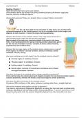
Reading guide for chapter 7 of Human Anatomy (9th Edition), by Marieb et al: "The Axial Skeleton". Used in the Applied Human Anatomy course at UC Irvine. Comes with bolded text answers and colored diagrams you can label.
Reading: Chapter 7
Name the bones that make up the axial skeleton.
Axial skeleton bones are found in the skull, vertebral column, and thoracic cage (ribs,
sternum, thoracic vertebrae) regions.
What is the hyoid bone? Where is it located? Why is it unique? What is its function?
It is the only bone that doesn’t articulate w/ other bones, but is tethered by
stylohyoid ligaments to the styloid process. It acts as a movable base for the tongue and
attaches to neck muscles -> moves the larynx during swallowing.
What are the functions of the vertebral column? How many bones are found in it;
what are these bones called and how are they separated?
Main support of the body axis: transmits weight of the trunk to the lower
limbs. It surrounds/protects the spinal cord and provides attachment points
for the ribs and neck/back muscles. Contains 26 bones called vertebrae,
separated by intervertebral discs.
What are the five major regions of the vertebral column (spinal column or spine)?
From superior to inferior: cervical vertebrae, thoracic vertebrae, lumbar
vertebrae, sacrum, coccyx.
How many vertebrae are in each region? Are the vertebrae fused or unfused?
● Cervical region: 7 vertebrae. Convex.
● Thoracic region: 12 vertebrae. Concave.
● Lumbar region: 5 vertebrae. Convex.
● Sacral region: consists of 5 fused sacral vertebrae and 3-5 fused
coccygeal vertebrae. Concave.
How does the shape of the vertebral column change superiorly to posteriorly?
From a lateral view, the column gives an S-shaped curvature: the cervical and lumbar regions
concave posteriorly, whereas the thoracic and sacral regions convex posteriorly.
How does this affect the function of the vertebral column?
Curvatures INCs resilience of the spine, acting as a spring rather than a straight rigid rod.
How are the vertebrae held together vertically by ligaments?
The anterior and posterior longitudinal ligaments run along the front and back vertebrae body
surfaces, attaching to both the body and the intervertebral discs. To prevent hyperflexion (head
forward, tearing spinal cord), the posterior longitudinal ligaments are only connected to the discs.
, Label the following ligaments:
Anterior longitudinal ligaments
Posterior longitudinal ligaments
What is an intervertebral disc? What is its function?
It is a cushion-like pad consisting of an inner sphere called the
nucleus pulposus and an outer collar called the anulus fibrosus(12
concentric rings). Their function is to absorb compressive stress
and limit expansion when the spine is compressed, respectively.
The rings also bind successive vertebrae together, resist tension in the
spine, and absorb compressive forces.
Describe the structure and function for each of the following general
features of a vertebra.
Body – weight-bearing region of the vertebrae.
Vertebral arch (composed of pedicles and laminae) –
consists of 2 pedicles and 2 laminae, forming the
posterior portion of the vertebra. Encloses/protects
the spinal cord and spinal nerves in the...
Vertebral foramen – hole created by the body and
vertebral arch. Successive vertebral foramina form the
vertebral canal, where the spinal cord and nerve roots
pass.
Spinous process – attachment site for muscles and
ligaments that move/stabilize the vertebral column.
Transverse process – projects laterally b/w the pedicle-
lamina junction. Attachment sites for muscles and
ligaments.
Transverse costal facet(2)-- facets on the transverse
processes that articulate w/ the tubercles of ribs. *Not
seen on T11-12.
Inferior articular process and facet – inferior process
joins w/ superior process of the vertebra immediately
inferior to it. Join successive vertebrae (along w/
discs).
Superior articular process and facet – ^
Intervertebral foramina – from a lateral view, the inferior and superior borders of pedicle form
opening s b/w the adjacent vertebrae. Allow nerves from the spinal cord to pass through.
Review Table 7.2 and be able to identify the general features and describe the structure and function
of the three main types of vertebrae.
Below are some questions that relate to some of the special characteristics of the regions of the
vertebral column.
What are the special features of the atlas (C1)? What is its function?
Atlas lacks a body/spinous process. Instead it is a ring: anterior and posterior arches, plus
lateral masses on each side. Atlas + occipital condyles = joint that allows for flexion and
extension of the head on the neck. “Yes”
What are the special features of the axis (C2)? What is the dens? What is its function?
The benefits of buying summaries with Stuvia:
Guaranteed quality through customer reviews
Stuvia customers have reviewed more than 700,000 summaries. This how you know that you are buying the best documents.
Quick and easy check-out
You can quickly pay through credit card or Stuvia-credit for the summaries. There is no membership needed.
Focus on what matters
Your fellow students write the study notes themselves, which is why the documents are always reliable and up-to-date. This ensures you quickly get to the core!
Frequently asked questions
What do I get when I buy this document?
You get a PDF, available immediately after your purchase. The purchased document is accessible anytime, anywhere and indefinitely through your profile.
Satisfaction guarantee: how does it work?
Our satisfaction guarantee ensures that you always find a study document that suits you well. You fill out a form, and our customer service team takes care of the rest.
Who am I buying these notes from?
Stuvia is a marketplace, so you are not buying this document from us, but from seller PrinceAlixD. Stuvia facilitates payment to the seller.
Will I be stuck with a subscription?
No, you only buy these notes for $5.49. You're not tied to anything after your purchase.