UCI BioSci D170, Williams Respiration
Reading: Chapter 22
What are the four processes of respiration?
Pulmonary ventilation (breathing), external respiration (O2-rich blood), transport of
respiratory gasses, and internal respiration (O2-poor blood).
What are the organs of the respiratory system?
Upper respiratory tract: nose, nasal cavity, paranasal sinuses, pharynx, larynx.
Lower respiratory tract: trachea, bronchi, lungs.
What is the difference between the conducting and respiratory zones?
The conducting zones include respiratory passageways that carry (filter, humidify, warm)
air to gas exchange sites. The respiratory zone is the actual site of gas exchange.
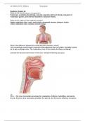
Describe the structure and function of the nose, noting the following structures.
Nose – The nose 1) provides an airway for respiration, 2) filters, humidifies, and warms
the air, 3) serves as a resonating chamber for speech, and 4) houses olfactory receptors.
, UCI BioSci D170, Williams Respiration
External nose – consists of the frontal, nasal, maxillary bones, and hyaline
cartilage. Septal cartilage forms the dorsum nasi; alar cartilage forms the apex (tip
of the nose); fibrous CT forms the ala (sides of nose).
Nasal cavity – posterior cavity divided into the right and left halves by the septum
(ethmoid’s perpendicular plate/vomer). Continuous w/ the pharynx through the
posterior nasal apertures.
Nares – external nares are the nostrils; internal nares are the...
Posterior nasal apertures – connects the nasal cavity to the nasopharynx, posteriorly.
Palate – forms the floor of the nasal cavity. -> Keeps food out of airways. Consists of the
har plate made of the palatine bones and palatine process, and the muscular soft palate.
Vestibule – superior to the nostrils, within the flared wings of the external nose, and lined
w/ nose hairs that filter large particles from the inspired air.
Respiratory mucosa – a mucous membrane that lines the vast majority of the respiratory
passageway. Consists of pseudostratified ciliated columnar epithelium containing goblet
cells; sitting on top of lamina propria containing mucous and serous (digestive enzyme)
cells. -> Secretes mucus that digests/destroys bacteria, filtering the inhaled air.
FUN FACT: a sneeze is stimulated when irritating particles contact the nerve endings
supplying the nasal mucosa.
Nasal conchae – scroll-like structures that project medially from each lateral wall:
superior, middle, and inferior conchae. Increases the surface area of the nasal mucosa.
Paranasal sinuses – ring of air-filled cavities surrounding the nasal cavity. Opens/mucus
drains into the nasal cavity, lined w/ respiratory mucosa, and helps warm/moisten
inhaled air.
Describe the structure and function of the pharynx, noting the following structures.
It is a funnel-shaped passageway for both food and air; walls consist of skeletal muscle.
Nasopharynx – superior division of the pharynx that serves as a passageway.
Uvula – pendulum on the soft palate that helps close off the nasopharynx during
swallowing.
Pharyngeal tonsil (adenoids)– lymphoid organs that destroy pathogens entering the
nasopharynx. A part of the posterior nasopharyngeal wall.
Oropharynx – extends inferiorly from the soft palate to the epiglottis; allowing swallowed
food and inhaled air to pass. Lining changes to stratified squamous epithelium ->
accommodates INCed friction and “chemical trauma” of food.
Laryngopharynx – serves as a passageway for food and air, lined w/ stratified squamous
epithelium. Posterior to the larynx and continuous w/ the esophagus.
Describe the structure and function of the larynx, noting the following structures.
Voicebox that attaches to the hyoid bone and opens into the laryngopharynx; continuous
w/ the trachea. 1) Produces vocalization, 2) provides an open airway, and 3) routes air
and food into the proper channels.
Thyroid cartilage – large, shield-shaped cartilage that covers the majority of the larynx.
Resembles an open book.
Laryngeal prominence – “spin of the open book.” Adam’s apple.
Cricoid cartilage – laryngeal cartilage inferior to the thyroid cartilage; forms a complete
ring on top of the trachea.
Epiglottis – leaf-shaped cartilage composed of elastic cartilage and covered by mucosa.
Attaches to the posterior aspect of the tongue and the internal aspect of the thyroid
cartilage. Covers the laryngeal inlet -> keeps food out of lower respiratory tubes.
Vocal folds (true vocal cords) – pair of mucosal folds formed by the vocal ligaments.
Exhaled air causes these folds to vibrate/clap together, producing speech.


