Summary Advanced Immunology Janeway test 5) Antigen presentation and immune regulation through B T cell interactions
0 purchase
Course
Advanced Immunology (5234ADIM6Y)
Institution
Universiteit Van Amsterdam (UvA)
Book
Janeway\'s Immunobiology
This is a small summary for the course advanced immunology from the master biomedical sciences at the UvA. It includes all the information you need for one of the 9 Janeway tests during this course. Look out for the bundle, because that's a lot cheaper!
Antigen presentation and immune regulation through B/
T cell interactions – Marieke van Ham
6-1
Processing & presentation antigens to T cells has two purposes:
Inducing development armed effector T cells.
Triggering effector functions.
Nearly all somatic cells (except red blood cells) have MHC I. CD8 T cell/cytotoxic T cell can recognise
these and kill them if something is wrong. MHC II is only expressed on cells from the immune system.
CD4 T cells can recognise this and become effector T cells (several subsets).
Direct presentation: Antigens from the cytosol are transported into the ER and loaded into MHC I
molecules. Antigens are from viruses and intracellular bacteria that replicate in cytosol. They are
able to replicate inside intracellular vesicles and do not get broken down by digestive enzymes.
Direct presentation: Extracellular antigens are presented into MHC II molecules after APCs have
taken them up via their endocytotic vesicles using specific cell surface receptors. B cells &
macrophages present antigens to recruit help from CD4 T cells.
Peptides derived from cytosolic proteins can also be
presented into MHC II via autophagy. Self is presented,
makes sure there is tolerance.
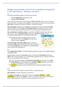
Cross presentation: Extracellular antigens (taken up by DC) are
presented into MHC I to be recognised by CD8 T cells (activation of T
cell is called cross priming). This is needed to for example eliminate
viruses that only infect epithelial cells.
6-3
The 2 peptide chains of MHC I are translocated during synthesis into the lumen of the ER folded &
assembled together.
Peptide/antigen transport into microsomes (vesicles of ER) requires ATP hydrolysis, delivered by
TAP1/2. TAP transports peptides of 8-16 aa (preferably hydrophobic/basic at C terminus) into the ER
before it can be loaded into MHC I molecules.
In the cytosol these peptides are protected from degradation by chaperones such as TRiC.
Amino terminus of too long peptides can be trimmed by ERAAP. Important, mice deficient
for ERAAP immunogenic for T cells from wild type mice.
IFN-gamma increases expression of ERAAP & other components of antigen-processing
pathway.
6-4
Newly synthesised MHC I molecules are held in the ER in a partly folded state.
MHC I deficiency: Less MHC I molecules on cell surfaces.
The folding and assembly of complete MHC I depends on association alfa chain with beta2
microglobulin and then with the peptide. It involves a lot of proteins with chaperone like functions.
When a peptide binds MHC I released from ER & transported to cell surface.
, MHC I alfa chains that enter ER bind calnexin beta2 microglobulin binds to alfa chain
dissociation from calnexin MHC I alfa:beta microglobulin binds to peptide loading complex (PLC,
including calreticulin, a chaperone, tapasin ERp57 & TAP). Tapasin forms a bridge between MHC I
molecules and TAP waiting for peptide from cytosol ERp57 and calreticulin bind other
glycoproteins, TAP is the final component of PLC and delivers peptides to partly folded MHC I.
ERp57 may have a role in
breaking and re-forming the
disulphide bond in the MHC I
alfa domain during peptide
loading.
PLC maintains the MHC I in a state that is
receptive for peptide binding and
mediates the exchange of low affinity
peptides to higher affinity peptides (by
tapasin, peptide editing). Most of the
peptides transported by TAP do not bind
MHC I and are transported back to the
cytosol by Sec61 (ATP dependent).
ERAD: Endoplasmic reticulum associated protein degradation. Several general cellular pathways
that involve the recognition and delivery of misfolded proteins to a retrotranslocation complex that
unfolds and translocates the proteins across the membrane of the ER to the cytosol. Proteins are
ubiquitinated during the process and are targeted to the proteasome (happens when TAP gene is not
functional).
In uninfected cells: Self peptides fill peptide binding groove of MHC I go to surface (only happens
when in excess, they wait because viral peptides must be immediately expressed).
6-5
Viruses have a different tropism, not all viruses infect DCs chance that these antigens are never
presented by DCs cross presentation.
Cross presentation is most efficiently performed by certain subsets of DCs. These DCs require
transcription factor BATF3 for development. They also uniquely express the chemokine receptor
XCR1.
In lymphoid tissues (spleen) they express CD8alfa molecule on the cell surface. Migratory
DCs capable of cross presentation are identified by expression of AlfaE integrin (CD103).
There may be several pathways for cross presentation.
There might be a direct pathway where PLC is transported from the ER to endosomal
compartments exogenous antigens into MHC I.
Another pathway may involve IRGM3 (induced by IFN-gamma). IRGM3 interacts with ADRP
in the ER and regulates generation of lipid bodies (storage of lipids, originate from ER
probably).
6-6
MHC II binds peptides generated in intracellular vesicles of DCs, macrophages and B cells to present
these to CD4 T cells. Some pathogens, like leishmania and mycobacteria (leprosy & tuberculosis) can
The benefits of buying summaries with Stuvia:
Guaranteed quality through customer reviews
Stuvia customers have reviewed more than 700,000 summaries. This how you know that you are buying the best documents.
Quick and easy check-out
You can quickly pay through credit card or Stuvia-credit for the summaries. There is no membership needed.
Focus on what matters
Your fellow students write the study notes themselves, which is why the documents are always reliable and up-to-date. This ensures you quickly get to the core!
Frequently asked questions
What do I get when I buy this document?
You get a PDF, available immediately after your purchase. The purchased document is accessible anytime, anywhere and indefinitely through your profile.
Satisfaction guarantee: how does it work?
Our satisfaction guarantee ensures that you always find a study document that suits you well. You fill out a form, and our customer service team takes care of the rest.
Who am I buying these notes from?
Stuvia is a marketplace, so you are not buying this document from us, but from seller nadinevankleef. Stuvia facilitates payment to the seller.
Will I be stuck with a subscription?
No, you only buy these notes for $5.90. You're not tied to anything after your purchase.