100% satisfaction guarantee Immediately available after payment Both online and in PDF No strings attached
logo-home
Adult Health Exam 3 $8.49
Add to cart

Class notes

Adult Health Exam 3

 0 view  0 purchase

Study guide for Exam 3

Preview 4 out of 34  pages

  • October 29, 2024
  • 34
  • 2023/2024
  • Class notes
  • Shotton & dercher
  • All classes
All documents for this subject (4)
avatar-seller
madisonbarton00
Adult Health Exam 3
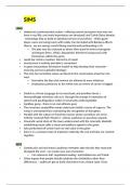
GI Changes with Aging – Stomach
Atrophy of Gastric Mucosa
Decrease in hydrochloric acid levels
- Decreased absorption of iron and vitamin B12
- Proliferation of bacteria
- Atrophic gastritis occurs as a consequence of bacterial overgrowth
Encourage bland foods & iron and B12
Assess epigastric pain


GI Changes with Aging – Intestine
Peristalsis decreases
Nerve impulses are dulled
Decreased sensation to defecate can result in postponement of bowel movements
- Leads to constipation and impaction
high fiber a fluid
activity !


GI Changes with Aging – Pancreas
Distension and dilation of pancreatic ducts
Calcification of pancreatic vessels occurs with a decrease in lipase production
- Decreased lipase level results in decreased fat absorption and digestion
- Excess fat in the feces (steatorrhea)occurs because of decreased fat digestion
small ,
frequent meals


GI Changes with Aging – Liver
Decrease in the number and size of hepatic cells and increase in fibrous tissue
- Leads to decreased protein synthesis and changes in liver enzymes
- Depresses drug metabolism – leads to accumulation of drugs, possibly to toxic levels
Assess AE of meds /toxicity ?)


Assessment – History and Physical Assessment
Patient history
Nutrition history
- Diet and food allergies
IMPORTANT:

- Anorexia and N/V
- Changes in taste
- Pain or difficulty swallowing
- Abdominal pain or discomfort with eating
- Dyspepsia – indigestion or heartburn
- Unintentional weight loss
- Alcohol and caffeine consumption
Family history and genetic risk
Current health problems
- Change in bowel habits
- Unintentional weight gain or loss
- Pain
- Changes in the skin due toalterations to


o
Discoloration or rashes, itching, jaundice, increased bruising, increased tendency to bleed
Physical assessment of abdomen

, - Inspection, Auscultation, Light Palpation, Percussion
- If appendicitis or an abdominal aneurysm is suspected, palpation is not done
Psychosocial assessment
- Stress can exacerbate some gastrointestinal disorders

Assessment – Labs
Liver Function Tests (liver enzymes)
- Alanine aminotransferase-ALT (4-36 units/L)
o Increased values may indicate liver disease, hepatitis, cirrhosis
- Aspartate aminotransferase–AST (0-35 units/L)
e o Increased values may indicate liver disease, hepatitis, cirrhosis
- Alkaline phosphatase-ALK (30-120 units/L)
o Increased values may indicate cirrhosis, biliary obstruction, liver tumor
yellow
tone
Bilirubin (0.3-1.0 mg/dL) – Increased values may indicate hemolysis, biliary obstruction, hepatic damage
Albumin (3.5-5) – Decreased values may indicate hepatic disease
Ammonia (10-80 mg/dL) – Increased values may indicate hepatic disease such as cirrhosis
colon
cancer, Ca 19-9 and CEA – Evaluated to diagnose cancer and could be increased in benign GI conditions
Serum amylase (30-220 units/L) – Increased values may indicate acute pancreatitis
Serum lipase (0-160 units/L) – Increased values may indicate acute pancreatitis
Prothrombin time (PT) (11-12.5 sec) – Useful in evaluating clotting, if elevated could indicate hepatic issue
Electrolytes
- Calcium (9-10.5) – Decreased values may indicate malabsorption, kidney failure, acute pancreatitis
- Potassium (3.5-5) – Decreased values may indicate vomiting, gastric suctioning, diarrhea, drainage from
intestinal fistulas
CBC – Low Hbg/Hct could indicate anemia with GI bleeding; elevated WBC could indicate infection
Stool – Annual guaiac heme fecal occult blood test (gFOBT) or fecal immunochemical test (FIT) to detect colorectal
cancer
- Ova and parasites – aid in diagnosis of parasitic infection, fecal fats, cytotoxic assay or culture

Assessment – Diagnostic Tests
Imaging Tests:
- Abdominal x-ray – can identify tumors, strictures, and obstructions
- Acute abdominal series – includes chest x-ray, supine and upright abdominal x-ray
- Abdominal computerized tomography (CT)
- Abdominal magnetic resonance imaging (MRI)
- Upper GI series (Barium Swallow) – X-ray from mouth to duodenojejunal junctions with use of barium -
risk for

FLUIDS
constipation

GIVE
- Small bowel follow-through – extension of the upper GI x-ray with use of barium
- Barium enema – X-ray of large intestine with use of barium
- Percutaneous transhepatic cholangiography (PTC) – examines biliary duct system using iodine dye
- Magnetic resonance cholangiopancreatography (MRCP)
Esophagogastroduodenoscopy (EGD)
- Visual exam of the esophagus, stomach, duodenum with use of fiberoptic scope
- Preparation: NPO for 6-8 hours and avoid anticoagulants, aspirin, NSAIDS several days before procedure
- Procedure: Moderate sedation and lasts about 20-30 minutes
- Post procedure:
o Keep patient NPO until gag reflex returns

o Priority care includes preventing aspiration and assess for any bleeding or pain that could
indicate perforation
Endoscopic retrograde cholangiopancreatography (ERCP)
- Visual and radiographic exam of the liver, gallbladder, bile ducts, and pancreas

, - Use radiopaque dye
- Used to diagnose obstruction as well as treat obstructions
- Preparation: NPO for 6-8 hours and typically avoid anticoagulants as determined by provider
- Procedure: Moderate sedation and lasts 30 minutes to 2 hours
- Post procedure:
o Keep patient NPO until gag reflex returns

o Priority care includes preventing aspiration and assess for any bleeding or pain that could
indicate perforation
o Assess for gallbladder inflammation and pancreatitis- severe abdominal pain, nausea and
vomiting, fever, and elevated lipase
Small bowel endoscopy (enteroscopy)
- Provides a visual view of the small intestine
- Used to evaluate and locate source of GI bleeding
- Preparation:
o NPO except water for 8-10 hours then complete NPO for 2 hours before swallowing capsule
-


- Procedure:
o Sensors are placed on abdomen and patient wears a data recorder

o Patient swallows the capsule endoscope and can resume normal activity

o Patient may eat 4 hours after swallowing the capsule

o Procedure lasts 8 hours
- Post procedure:
o Explain to the patient that the capsule endoscope is excreted naturally and will be seen in the
stool
Colonoscopy
- Endoscopic exam of the entire large intestine – can be used to visually diagnose, biopsy and treat
- Baseline test should be done at age 50 and every 10 years
- Preparation:
o Clear liquids the day before

o NPO 4-6 hours prior

o Avoid aspirin, anticoagulants, and antiplatelet drugs for
several days before
o Adequate bowel cleansing is essential – follow provider orders for oral and rectal preparation;
Patient should be passing clear liquid prior to procedure
- Procedure: Moderate sedation and procedure lasts 30-60 minutes
- Post procedure:
o Observe for signs of perforation (severe pain) and hemorrhage

o Feelings of fullness and cramping are expected

o Fluids are permitted after the patient passes flatus to indicate that peristalsis has returned


Stomatitis
Inflammation within the oral cavity
- Painful single or multiple ulcerations that appear as inflammation and erosion of the protective lining of the
mouth; sores cause pain and open areas place the person at risk for bleeding and infection
- Mild erythema (redness) may respond to topical treatments

, - Extensive stomatitis may require treatment with systemic analgesics or medications
Causes:
- Infection – bacteria and viruses have a role in recurrent stomatitis
- Allergies – certain foods, such as coffee, potatoes, cheese, nuts, citrus fruits, and gluten may trigger allergic
responses that cause aphthous ulcers
- Vitamin deficiency – B vitamins, folate, zinc, iron
- Systemic disease and immunosuppression
- Irritants – tobacco and alcohol
Assessment:
- History of recent infections
- Nutritional changes
- Oral hygiene habits or oral trauma
- Stress
- Drug history
- If lesions are seen along the pharynx and the patient reports painful swallowing, the lesions might extend
down the esophagus
Interventions:
- Remove dentures
- Encourage or provide oral hygiene after each meal and as often as needed
- Increase oral care to every 2 hours or more
- Use a soft toothbrush or gauze
- Encourage frequent rinsing of the mouth with warm saline or baking soda solution
- Avoid commercial mouthwashes, specifically ones with alcohol and lemon/glycerin
- Assist with food choices
o Soft, bland, and nonacidic foods can decrease irritation; cool food can be soothing
Drug therapy:
- Antimicrobials for control of infection
o Ex. Tetracycline, Minocycline, chlorhexidine mouthwashes
- Herpes simplex
o IV Acyclovir (Zovirax) if severe or with immunocompromised patient; can be given oral or
topical
- Fungal infections/Yeast
o Nystatin (Mycostatin) swish/swallow
- Pain control
o Anesthetics; ex. Orabase, Anbesol, Campho-Phenique


Oral Cancer
Squamous cell carcinoma -
most common


- Oral lesions appear as red, raised, eroded areas on the lips, tongue, buccal mucosa, and oropharynx
- 90% of oral cancers – major risk factors are increasing age, tobacco use, alcohol use
Basal cell carcinoma
- Oral lesion is asymptomatic and resembles a raised scab – occurs primarily on the lips
- Lesion evolves into a characteristic ulcer with a raised, pearly border
- Major risk factor is excessive sunlight exposure
Kaposi’s Sarcoma
- Malignant lesion in blood vessels – usually painless and appears as a raised, purple nodule or plaque
- Most common site is the hard palate but can be found on the gums, tongue, or tonsils
- Most often associated with AIDS
Clinical Manifestations
- Bleeding from the mouth, poor appetite, difficulty chewing, difficulty swallowing, poor nutritional status and

The benefits of buying summaries with Stuvia:

Guaranteed quality through customer reviews

Guaranteed quality through customer reviews

Stuvia customers have reviewed more than 700,000 summaries. This how you know that you are buying the best documents.

Quick and easy check-out

Quick and easy check-out

You can quickly pay through credit card or Stuvia-credit for the summaries. There is no membership needed.

Focus on what matters

Focus on what matters

Your fellow students write the study notes themselves, which is why the documents are always reliable and up-to-date. This ensures you quickly get to the core!

Frequently asked questions

What do I get when I buy this document?

You get a PDF, available immediately after your purchase. The purchased document is accessible anytime, anywhere and indefinitely through your profile.

Satisfaction guarantee: how does it work?

Our satisfaction guarantee ensures that you always find a study document that suits you well. You fill out a form, and our customer service team takes care of the rest.

Who am I buying these notes from?

Stuvia is a marketplace, so you are not buying this document from us, but from seller madisonbarton00. Stuvia facilitates payment to the seller.

Will I be stuck with a subscription?

No, you only buy these notes for $8.49. You're not tied to anything after your purchase.

Can Stuvia be trusted?

4.6 stars on Google & Trustpilot (+1000 reviews)

52928 documents were sold in the last 30 days

Founded in 2010, the go-to place to buy study notes for 14 years now

Start selling
$8.49
  • (0)
Add to cart
Added