Detailed English summary for the course 3.6C Neuropsychology with all the important information for the exam. Contains information about all 4 problems and the tutorials, including visuals.
Although quite long and exhaustive, the summary contains all the information you need for the exam, simplif...
Kolb & Wishaw – Chapter 14: The Parietal Lobes
• The parietal cortex processes and integrates somatosensory & visual information, especially with
regard to controlling movement.
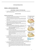
Parietal Lobe Anatomy
Subdivisions of the Parietal Cortex
• Parietal region of the cerebral cortex lies between the frontal & occipital lobes,
underlying the parietal bone at the roof of the skull.
• This area is demarcated anteriorly by the central fissure, ventrally by the lateral
(Sylvian) fissure, dorsally by the cingulate gyrus, and posteriorly by the parieto-
occipital sulcus.
• Principle regions of the parietal lobe include the postcentral gyrus (Brodmann’s
areas 3-1-2), superior parietal lobule (areas 5 & 7), parietal operculum (area 43),
supramarginal gyrus (area 40), and angular gyrus (area 39).
• Together, the supramarginal gyrus & angular gyrus are often referred to as the
inferior parietal lobe.
• Parietal lobe can be divided into 2 functional zones --> an anterior zone including
areas 3-1-2 and 43 & a posterior zone that includes the remaining areas.
o Anterior zone is the somatosensory cortex & Posterior zone is called the
posterior parietal cortex.
• Over the course of human evolution, parietal lobes have undergone a major
expansion, largely in the inferior region.
o This size increase makes comparisons of various areas in the human brain
with those in the monkey brain confusing, especially because while
Brodmann identified areas 39 & 40 in the human brain, he failed to
identify them in the monkey.
o Whether monkeys actually have regions homologous to these areas is
debatable.
• On Constantin van Economo’s (another anatomist) maps, in which parietal areas
are called PA (parietal area A), PB, and so forth, are 3 posterior parietal areas (PE,
PF, PG) that von Economo described in both humans & monkeys.
o Area PF is equivalent to Brodmann’s areas 43 and 40
plus part of area 7 and PE to area 5 and the remainder of area 7.
o Area PG is roughly equivalent to Brodmann’s areas 39 & 40.
§ These PG areas are primarily visual & include areas AIP, LIP, cIPS, and PRR.
• An area significantly expanded in the human brain appears to consist of the polymodal parts of area
PG & adjoining polymodal cortex in the superior temporal sulcus (STS).
o Polymodal cells receive inputs from more than one sensory modality.
o Those in PG respond to both somatosensory & visual inputs, whereas those in the STS (3rd
visual pathway) respond to various combinations of auditory, visual, and somatosensory
inputs.
1
, • Increased size of area PG & STS is especially interesting, because this region is anatomically
asymmetrical in the human brain.
o The asymmetry may be due to a much larger area PG (and possibly STS) on the right than on
the left.
o If PG has a visual function and is larger in humans, especially in the right hemisphere, then
we might expect unique visual symptoms after right parietal lesions --> This indeed is the
case.
o Note, however, that PG is also larger on the left in the human than in the monkey --> This
leads us to expect unique deficits in humans after left-hemisphere lesions, which is the case
as well.
• Specific parietal regions take part in the dorsal stream of visual processing, in particular the
intraparietal sulcus (cIPS) and the parietal reach regions (PRR).
• Regions in the intraparietal sulcus contribute to controlling saccadic eye movements (area LIP) and
visual control of object-directed grasping (AIP).
o Saccade: A series of involuntary, abrupt, and rapid small movements or jerks made by both
eyes simultaneously in changing the point of fixation.
• PRR has a role in visually guided grasping movements.
Connections of the Parietal Cortex
• Anterior parietal cortex makes rather straightforward connections.
• Projections from the primary somatosensory cortex extend to secondary somatosensory area PE,
which has a tactile recognition function, as well as to motor areas including the primary motor cortex
and the supplementary motor and premotor regions in the frontal lobes.
• Motor connections are important for providing sensory info about limb position in movement control.
• Although more than 100 inputs & outputs have been described for areas 5 & 7 in the monkey (PE, PF,
and PG), a few basic principles summarize the connections:
o 1) Area PE is basically somatosensory, receiving most of its connections from the primary
somatosensory cortex. PE’s cortical outputs are the primary motor cortex and to the
supplementary motor (SMA) and premotor regions, as well as to PF. Area PE therefore plays
some role in guiding movement by providing info about limb position.
o 2) Area PF has heavy input from the primary somatosensory cortex through area PE. PF also
receives inputs from the motor & premotor cortex and a small visual input through area PG.
PF’s efferent connections are similar to those of area PE, and these connections presumably
elaborate similar info for the motor systems.
o 3) Area PG receives more-complex connections including visual, somesthetic (skin
sensations) proprioceptive (internal stimuli), auditory, vestibular (balance), oculomotor (eye
movement), and cingulate (motivational?). Critchley first described area PG as the ‘parieto-
temporo-occipital crossroads’, which is apparent from the connectivity. Its function likely
corresponds to this intermodal mixing. Area PG is part of the dorsal stream that controls
spatially guided behavior with respect to visual & tactile info.
2
, o 4) The close relation between the posterior parietal connections & the prefrontal cortex are
apparent in the connections between the posterior parietal cortex (PG & PF) and the
dorsolateral prefrontal region. Additionally, the prefrontal and the posterior parietal regions
project to the same areas of the paralimbic cortex and the temporal cortex as well as to the
hippocampus and various subcortical regions. These connections emphasize a close
functional relation between the prefrontal and parietal cortices. This relation probably has
an important role in controlling spatially guided behavior.
Anatomy of the Dorsal Stream
• As originally conceived by Ungerleider & Mishkin, the dorsal stream from the occipital cortex to the
posterior parietal regions was conceived of as a ‘where’ pathway.
• Based on their studies, Milner & Goodale hypothesized the dorsal stream as a ‘how’ pathway.
• Since then, our knowledge of the dorsal stream’s organization & function has increased vastly, leading
Kravitz et al. to construct a new framework for it.
• Kravitz et al. identify 3 functional pathways leaving the posterior parietal region & traveling to the
premotor, prefrontal, and medial temporal regions (figure 14.3C).
o Parieto-premotor pathway is proposed as the principal ‘how’ pathway.
o Parieto-prefrontal pathway is proposed to have visuospatial functions, especially related to
visuospatial working memory.
o Parieto-medical temporal regions as well as indirectly via the posterior cingulate and
retrosplenial cortex, is proposed to have a role in spatial navigation.
o Thus, the posterior parietal cortex would contribute to the dorsal stream by participating in
nonconscious visuospatial behavior, that is, reaching for & grasping objects.
• Caution against seeing these pathways as entirely divergent --> The goal of all dorsal stream pathways
is to guide visuospatial behavior through motor output, so the parieto-prefrontal and parieto-
3
, mediotemporal pathways must eventually influence motor output, though more indirectly than does
the parieto-premotor pathway.
A Theory of Parietal-Lobe Function
• If we consider the anterior (somatosensory) & posterior (spatial) parietal zones functionally distinct,
we can identify 2 independent parietal-lobe contributions.
o Anterior zone processes somatic sensations & perceptions.
o Posterior zone specializes primarily in integrating sensory input from the somatic & visual
regions and to a lesser extent from other sensory regions, mostly for controlling movements
– reaching & grasping as well as whole-body movements in space.
§ Posterior parietal cortex also plays a significant role in mental imagery, especially
related to both object rotation & navigation through space.
• As we think about how the brain manages such tasks, an internal representation of the location of
different objects around us seems obvious, a sort of map in the brain of where things are.
o Furthermore, we assume that the map must be common to all of our senses, because we can
move without apparent effort from visual to auditory to tactile info.
o Parietal lobe plays a central role in creating this brain map.
• We take for granted that the world around us is as we perceive it and thus that the brain employs a
unified spatial map --> Real space must be mapped topographically in the brain because that’s how it
appears to us.
• However, few evidence supports the existence of such a map in the brain --> More likely is a series of
neural representations of space that vary in 2 ways:
o 1) Different representations serve different behavioral needs.
o 2) Spatial representations vary, from simple ones applicable to controlling simple
movements to abstract ones that may represent info such as topographic knowledge.
Behavioral Uses of Spatial Information
• Milner & Goodale emphasize that we need spatial info about the location of objects in the world,
both to direct actions at those objects & to assign meaning and significance to them.
o In this sense, spatial info is simply another property of visual info, much like form, motion,
and color.
o Just as form is coded in more than one way in visual processing, so is spatial info.
o The critical factor for both form & space lies in how the info is to be used.
• There are 2 basic types of form recognition; one for recognizing objects & the other for guiding
movements to objects --> We can think of spatial info in the same way.
• Object Recognition: The spatial info needed to determine relations between objects, independent of
what the individual’s behavior might be, is very different from the spatial info needed to guide eye,
head, or limb movements to objects.
o In the latter case, visuomotor control must be viewer-centered --> The object’s location & its
local orientation and motion must be determined relative to the viewer.
o Because the eyes, head, limbs, and body are constantly moving, computations about
orientation, motion, and location must take place every time we wish to undertake an
action.
o Details of an object’s characteristics, such as color, are irrelevant to visuomotor guidance of
viewer-centered movements --> A detailed visual representation isn’t needed to guide hand
action.
o Brain operates on a ‘need-to-know’ bias.
§ Having too much info may be counterproductive for any given system.
4
Los beneficios de comprar resúmenes en Stuvia estan en línea:
Garantiza la calidad de los comentarios
Compradores de Stuvia evaluaron más de 700.000 resúmenes. Así estas seguro que compras los mejores documentos!
Compra fácil y rápido
Puedes pagar rápidamente y en una vez con iDeal, tarjeta de crédito o con tu crédito de Stuvia. Sin tener que hacerte miembro.
Enfócate en lo más importante
Tus compañeros escriben los resúmenes. Por eso tienes la seguridad que tienes un resumen actual y confiable.
Así llegas a la conclusión rapidamente!
Preguntas frecuentes
What do I get when I buy this document?
You get a PDF, available immediately after your purchase. The purchased document is accessible anytime, anywhere and indefinitely through your profile.
100% de satisfacción garantizada: ¿Cómo funciona?
Nuestra garantía de satisfacción le asegura que siempre encontrará un documento de estudio a tu medida. Tu rellenas un formulario y nuestro equipo de atención al cliente se encarga del resto.
Who am I buying this summary from?
Stuvia is a marketplace, so you are not buying this document from us, but from seller dogaultanir. Stuvia facilitates payment to the seller.
Will I be stuck with a subscription?
No, you only buy this summary for $18.97. You're not tied to anything after your purchase.