Comprehensive lecture notes for the Protein processing, traffiking and turnover module covered in MCB3025F, speficially for sections 1 and 2 covering Protein folding and disease (Alzheimer’s Disease as a model) and Post-translational modifications and Disease (Huntington’s Disease as a mode). T...
Protein Folding, Modification, Transport and Degradation
Section 1: Protein folding and disease (Alzheimer’s Disease as a model)
Part A: Protein folding
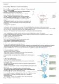
- in steps 1-3, DNA makes RNA (process of transcription) and
RNA makes protein (process of translation)
- after translation (step 3), the translated protein is not in its
mature stage and is unable to perform its role
- in order to become a functional protein, the newly translated
protein must progress through steps 4-7 to complete its life cycle
- not that proteins after protein PTM are rerouted to step 6 for
transport
- during protein transport, note that a fraction of p proteins do not
undergo PTM and proceed directly from step 4 to 6 (folding step
to transport)
- finally, protein degradation
- a functional protein is one that is in its mature 3D structure and performs its function at the correct location
- in an ideal world, we would like all these steps to function expertly to obtain all functional proteins however, this is not the
case and when any of these steps malfunction, this leads to human diseases
- hence, we will relate these steps to human diseases to understand its functional importance
- linear protein sequence is in the immature protein and protein folding leads to mature 3D structure
In order to understand how the folding step to reach its mature 3D structure occurs, are 6 questions we need to answer
1) where do newly synthesized proteins fold?
2) Why is protein folding important?
3) what is the protein folding process?
4) how does a protein know in what 3D conformation to fold?
5) how do newly synthesized proteins fold in vivo
6) what happens if protein folding mechanisms fail?
1) Where newly translated proteins fold in the cell
- in eukaryotic cells, protein folding can occur in the cytoplasm, the mitochondria and in the rough endoplasmic reticulum
- all proteins being their translation in the cytoplasm on free ribosomes (free because they are floating around in the
cytoplasm) but from there, can move into the mitochondria or rough ER to complete their folding process
2) Why protein folding is important
- protein structure is critical to function
- helps protein to reach its biologically active/native 3D conformation
- stabilizes the protein by minimizing free energy
- regulates activity and signaling (creates binding sites for other ligands to
bind)
3) The protein folding process
- refers to the 4 levels of protein structure
- first is the primary structure which refers to the sequence of amino acids
making up the chain
- the types of amino acids and their position are important in determining the
3D shape of the protein
- next level is the secondary structure, commonly referred to as beta sheets and
alpha helices
- these are formed by hydrogen bonding in the peptide backbone
- the third level is the tertiary structure which is the 3D conformation and is
the biologically active or native form of the protein
- it is formed by side chain interactions of the amino acids making up the chain
- most proteins complete the folding process at the 3rd level but some proteins
that are made up of multiple polypeptide chains will go on to form a
quaternary structure
- the quaternary structure come together to form a functional protein
Recap: Disulphide bonds in 3D strucure
,- covalend bond formed between the SH-groups (sulfhydryl groups) of two cysteine residues (R-S-S-R) which significantly
stabilizes the 3D stcucture
- this reaction is catalysed by a specufuc enzyme called protein disulphide isomerase
- both the tertiary, and in some cases, the quaternary structure, is the biologically active form of the proteim and both of
these structures are stabilised by disulphide bonds
- take not that at the startm the cysteine residues have the sulfhydryl side chain and in a sequence view, take note of the
methionine as the first amino acid making up the protein chain
- have a cystiene residue at position 6 and position 15 which form a disulphide bonds to form a loop (a fold) in the protein
4) How a protein knows in what 3D conformation to fold
- Christian Anfinsen performed a very simple experiment using Ribonuclease A
(RNaseA) to study protein folding
- RNaseA degrades RNA in a cell and this protein is 124 amino acids in length which
has 4 disulphide bonds
- Anfinsen used Ribonuclease A together with urea and beta- mercaptoethanol (ME)
- urea breaks hydrogen bonds in a protein and mercaptoethanol breaks down disulphide
bonds in a protein
Anfinsen’s experiment
- he performed in three different stages
- in stage 1, he treated native RNaseA with mercaptoethanol and urea and found that the
treatment with both of these chemicals completely denatured the protein back to its linear sequence (primary structure) and
disulphide bonds broken
- in the same experiment, he then removed mercaptoethanol and urea and found that the protein spontaneously refolded
back into its biologically active 3D conformation and reformed the correct disulphide bonds
- this suggested to him that the primary sequence of amino acids making up the protein was enough for the protein to reform
into its native conformation
- in stage 2, he took denatured RNaseA and treated it with urea, but he completely removed mercaptoethanol
- found that an inactive or scrambled RNaseA was formed
- this was because the protein randomly formed disulphide bonds (formed
incorrect disulphide bonds)
- this suggested to him that the absence of the hydrogen bonds (because
of the presence of the urea) impaired the proteins ability to form correct
disulphide bonds
- in stage 3, he took the scrambled, inactive RNaseA and treated it with
trace amounts of mercaptoethanol, but he completely removed urea
- found that over time, the protein was able to fold into its biologically
active 3D conformation with the correct disulphide bonds
- possible because the trace amounts of beta-mercaptoethanol was
enough to break down the incorrect disulphide bonds allowing the protein
to try and form the correct disulphide bonds and to fold into its native
state that was thermodynamically most favorable and lowest in energy
(trial-and-error process to allow the protein to reach its final stage)
Conclusions
1. Information for forming the mature 3D structure of a protein is determined by its primary structure (amino acid sequence)
2. Folding proceeds in ordered states; the information in primary sequence drives hydrogen bonding in secondary structure
which then drives correct formation of disulphide bonds in the 3D state
3. proteins will fold to reach the thermodynamically most stable, favored state
5) how newly translated proteins fold in vivo
, - the main driving force behind protein folding is the hydrophobic effect and it is based on non-polar hydrophobic groups
clustering away from the aqueous solution
- protein folding occurs in a region of high protein concentration and hence folding proteins are very likely to aggregate
with each other through hydrophobic interaction
- to prevent this problem, protein folding is assisted by chaperones and chaperonins
- overall, chaperones and chaperonins assist protein folding by preventing non-specific hydrophobic interactions, providing
a chamber to isolate folding and stabilizing unfolded proteins during transport to subcellular organelle (by binding to the
hydrophobic groups on the polypeptide chains, preventing misfolding and aggregation as it is being transported)
Chaperones and chaperonins
- are found in all organisms and many of these are called heat shock proteins (HSP) because their expression was noted after
a cell was exposed to hear
- the number following the HSP usually denotes the molecular weight of the protein
- the chaperones bind to the proteins while they are still being synthesized by the ribosomes to prevent them from
misfolding, such that it enhances the formation of biologically active native 3D stage of the protein
- some proteins however go on to use a chaperonin to facilitate the folding process
- a chaperonin will function after a chaperone and in this case, the chaperonin forms an isolation chamber within which the
protein can fold away from other proteins to achieve the native state
- example of a protein that uses both chaperone and a chaperonin is actin
Structure of HSP70 (a chaperone)
- a very common chaperone is HSP70 found in the cytosol as
well as in the mitochondria
- another example is BIP (binding immunoglobulin protein) that
is round in the PR
- structurally, HSP70 has two major domains
- has an ATPase domain at the N terminal which binds and
hydrolyses ATP to ADP which proves the energy to power
binding and release of the target protein of HSP70
- at the C terminal, has the substrate binding domain and the
helical lid region
- the substrate binding domain contains a substrate hydrophobic
binding pocket onto which hydrophobic residues in the target
protein can bind
- the lip region provides a lid that clamps down over the
substrate biding pocket once a target protein has bound
Function of HSP70
- HSP70 functions in an open and closed format
- in panel 1, not interacting with any target protein and HSP is bound to ATP and assumes an open form (its hydrophobic
pocket within the substrate binding domain is open and available for hydrophobic residues within a target protein to bind)
- this binding of the target protein to the hydrophobic
pocket causes hydrolysis of ATP to ADP
- this hydrolysis provides energy for the lid region to
clamp down over the target protein, keeping the
protein bound to the HSP70
- in panel 3, exchange of ADP for ATP causes the lip
region to open, releasing the target protein from the
hydrophobic pocket, which at this stage could have
partially folded
Structure of HSP60 (a chaperonin)
Los beneficios de comprar resúmenes en Stuvia estan en línea:
Garantiza la calidad de los comentarios
Compradores de Stuvia evaluaron más de 700.000 resúmenes. Así estas seguro que compras los mejores documentos!
Compra fácil y rápido
Puedes pagar rápidamente y en una vez con iDeal, tarjeta de crédito o con tu crédito de Stuvia. Sin tener que hacerte miembro.
Enfócate en lo más importante
Tus compañeros escriben los resúmenes. Por eso tienes la seguridad que tienes un resumen actual y confiable.
Así llegas a la conclusión rapidamente!
Preguntas frecuentes
What do I get when I buy this document?
You get a PDF, available immediately after your purchase. The purchased document is accessible anytime, anywhere and indefinitely through your profile.
100% de satisfacción garantizada: ¿Cómo funciona?
Nuestra garantía de satisfacción le asegura que siempre encontrará un documento de estudio a tu medida. Tu rellenas un formulario y nuestro equipo de atención al cliente se encarga del resto.
Who am I buying this summary from?
Stuvia is a marketplace, so you are not buying this document from us, but from seller ggauntlett. Stuvia facilitates payment to the seller.
Will I be stuck with a subscription?
No, you only buy this summary for $2.96. You're not tied to anything after your purchase.