NEUROCOGNITION
LECTURE 1: Development of cognition over the lifespan
The nervous system consists of two main parts:
1. Central Nervous System (CNS): the brain and spinal cord.
2. Peripheral Nervous System (PNS): the autonomic and peripheral sensory and motor
system.
The brain has different types of neurons, categorized by their shape and function:
• Sensory neurons: collect information (afferent)
• Interneurons: make connections within the brain
• Motor neurons: give motor output (efferent)
The neurons communicate with each other through chemical
action potentials, that are triggered by a summation of
excitatory potentials. These are tresholded and non-
decremental. This means you can’t have a ‘little’ action
potential. There are differences in frequency, but not in size.
Glia cells: next to neurons, the brain also has different types of glia cells:
• Astrocytes: blood-brain barrier and structural support.
• Oligodendrocytes: what gives our cells the myelin to facilitate communication for
CNS neurons.
• Microglial cells: smaller, doing ‘household chores’ such as infection-fighting and
waste disposal.
• Ependymal cells: Create CSF
• Schwann cells: these cells are not in the brain but provide myelin for the peripheral
neurons (same as oligodendrocytes, but in the peripheral area).
White matter tracts: all the axons, with their myelin, are connected underneath the shell of the
cortex as white matter tracts. These connect different brain areas in a number of ways.
• Association fibers: connecting areas within a hemisphere.
• Commissural fibers: crossing to the other hemisphere, to the same (homotopic) or a
different place (heterotopic).
• Projection fibers: connect outwards, to subcortical regions / cerebellum / spinal cord.
These white matter tracts are similar between people, so we can recognize specific
connections and directions in the brain. When a particular tract is not visible/not working the
same, this could show that some connections are not working.
Grey matter = neuronal cell bodies and glia cells, situated on the outer layer of the
hemispheres and in subcortical nuclei.
White matter = neuronal axons and glia cells, organized in bundles that connects different
grey matter areas.
,The CNS consists of the following areas:
1. Forebrain (telencephalon): the hemispheres, the
corpus callosum and subcortical deep structures.
2. Diencephalon: thalamus, hypothalamus, pituitary gland
3. Midbrain (mesencephalon): top of brain stem, incl.
sensory and motor relay nuclei.
4. Hindbrain (metencephalon): pons, cerebellum,
medulla oblongata.
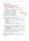
Thalamus: Basically, everything that goes in
or out, goes through the thalamus = the
connections go throughout the whole brain.
It is a relay station consisting of many
nuclei.
Image: the grey areas in the middle, are the
subcortical structures. The thalamus is not
really considered subcortical but could be
added.
Basal ganglia: the motor, associative (learning) and
reward circuit go through the basal ganglia. These
circuits are clearly related, but they are anatomically
separate.
- Some areas need to be inhibited, and some need
to be excited, which creates different loops in the
basal ganglia.
Limbic system: this is the emotional coloring of our
experiences. It consists of the cingulate cortex,
hippocampus, hypothalamus (because of this, the
hormone system is included), and amygdala.
- Emotional processing
- Fight/flight processes
The lobes of the brain:
- Frontal: movement, attention, reward, short-term
memory, planning, impulse control: creating
behavior, focused on the output. The non-motor
part of this lobe is the prefrontal cortex. This is
the most cognitive part of the brain.
- Parietal: sensory integration, association
processes, language, spatial processing, sense of
, touch, some visual processes. Important function of this lobe is the multiple maps of
body space. This means that you know where your body is without using visual input.
- Occipital: visual processes.
- Temporal: integration of multiple types of processing, memory, emotion association,
primary auditory areas. Wernicke’s area is located in this lobe.
o Medial temporal lobe: this part of the temporal lobe differs in function and is
typically described as a separate structure. The MTL is home to the
hippocampi and memory functioning. It includes the whole limbic system.
The brain of two hemispheres that are completely separated. The link between them is
provided by the corpus callosum, a large arch of white matter. While the left and right
hemispheres have some different functions, the corpus callosum has 100 million fibers,
constantly trafficking back and forth, which means integration of information from both sides.
Cross-lateralized = when I move my left arm, it is working in my right hemisphere.
Exceptions of this are:
- Language is usually left-lateralized, having non-verbal material on the right.
- Global perception is usually right-lateralized, local perception is on the left.
Gyri: bumps/ridges in the wrinkles
Sulcus: groove in the wrinkles, big ones are called
fissures.
CSF: Cerebrospinal fluid works as a cushion for the
brain, so it protects the brain. It circulates nutrients
and chemicals and removes waste. It runs in:
- Ventricles
- Subarachnoid space (space between the
brain and the layers)
- Venous sinus
CSF is created in the lateral ventricles by the
ependymal cells.
Naming conventions:
- Brodmann: histological. He looked at the structure of the cell.
These areas are about the tissue type/structure, but this doesn’t really
go along with the functioning. It only sometimes overlaps with
functionally distinct areas.
- Functional names: exact location can stay vague, assumes one
function per area.
- Relative locations: for example, ventromedial thalamic nucleus.
- Coordinate systems: This is the most precise way. For example, the
MNI coordinates, which are based on ‘standard brain’.
, Normal brain aging and cell
development:
- When the brain changes,
the dendritic spines are
being formed. This is
dependent on life stage,
long-term potentiation,
or depression, and
pruning of receptive
spines on the dendrites.
- Neurogenesis: The
creating of neurons. Only some brain areas are known to be able to grow new neurons.
- Apoptosis/pruning: the death of neurons/cell death. This is different from necrosis
(cell death due to external causes) because apoptosis is as a developmental process.
As we age, the brain changes:
- Cortical thinning: ventricles become bigger.
- Neuronal and synaptic loss: the brain shrinks.
- White matter lesions
- Inflammations
- Beta-amyloid plaques, neurofibrillary tangles:
degradation of cells
Brain functioning changes across age. Increased activation for the same tasks can be due to
reorganization or compensation. It is difficult to find out what the cause of the increased
activation is.
Structural plasticity can happen through experience: when you practice something really
often, this may make differences in brain activity. This means that long-term training can lead
to brain change, depending on aspects of training and sensitive developmental periods.
- Image: string players have one hand with highly developed motor skills (more
wrinkles), pianists have two.
Brain functioning can reorganize dynamically: for example, if the part of the brain isn’t used
(for example vision areas for blind people), functions can migrate to these unused brain
areas.
Modular concept: each area has its own function.
Network concept each function is implemented by a combination of regions.
- Some areas are necessary for specific functions, but many deficits arise due to
connections being broken.
Changes in the brain due to damage:
- Normal aging: atrophy