Enrico Tiepolo
Respiratory system
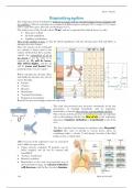
The respiratory system is designed to bring air in contact with an extended surface for gas exchange with
the capillaries. The air we breathe in is composed of different gases: nitrogen (78%), oxygen (21%), argon
(0.9%), and other gases, like CO2 and hydrogen (0.1%).
The surface area of the alveoli is about 70 m2, and air is separated from blood, there, by only:
• Thin layer of fluid.
• Alveolar epithelium.
• Capillary endothelium.
Flow in the capillary is slow, so that the blood equilibrates with the alveolar gases (O2 and CO2, as
nitrogen does not change).
Since the amount of air exchanged
in a minute is about equal to the
volume of blood that flows (cardiac
output), the composition of air in
the alveoli is not the same as in
external air: O2 will be lower,
CO2 will be higher, and the air
will be warm and humid (high
partial pressure of water vapor).
Before entering the alveolar ducts
and finally the alveolar sacs, the air
moves through:
• Larynx
• Trachea
• Bronchi
• Bronchioles
• Terminal bronchioles
• Respiratory bronchioles
Bronchi become increasingly numerous and small.
The total cross-sectional area increases enormously in the late
generations of terminal bronchioles and in respiratory
bronchioles, so that the flow velocity correspondingly declines.
Flow velocity and the presence of ramifications play an important
role in determining whether the flow of air in the respiratory
apparatus is laminar, turbulent or transitional (a mix of the
two).
The flow switches from laminar to turbulent when Reynolds’
number [the ratio of inertial to viscous forces, given by
combining radius r, velocity V, and density/viscosity of the fluid
Y/7] exceeds a critical value.
Different areas of the respiratory tract are associated
with a different types of flow:
• Upper airways: turbulent à particles can be
better trapped, and air can be warmed and
humidified.
• Trachea: transitional
• Bronchi: turbulent
• Bronchioles: as the total cross-sectional area of
the bronchioles is huge, the velocity of the flow
will decrease, and the flow becomes laminar.
149 Body At Work II
, Enrico Tiepolo
When it exceeds a certain value, flow will become turbulent, increasing the contact surface of air with
lining of the respiratory tract, and particles that may be present in the air we breathe will tend to hit the
walls and be trapped by mucus. This is one function of the turbinate bones in the nose, which create
a turbulent flow and therefore should avoid any big particle entering the airways.
Any particles that manage to enter the airways will however tend to deposit at different depths in the
bronchial tree, depending on their size; the smallest ones will deposit in respiratory bronchioles where
the flow is very slow and changes direction.
Only particles smaller than 0.5 µm will manage to enter in the alveoli and here they are either expelled
when we breath out, or they can be trapped and adhere to the walls, where they will be cleared by
macrophages (causing inflammation).
Respiratory mechanics
The lungs have an elastic structure; if isolated, they tend to shrink. In situ, in the thorax, they
cannot shrink because they are attached to the thoracic wall through the visceral and parietal pleural
membranes, but they will pull on the thorax forcing it to a smaller volume than the one it would
have a passive empty cage.
In simple words, the lungs are pulling on the thorax to shrink, while the thorax is pulling on
the lungs to expand. Since they are both attached to each other, whichever pulls more is going to win
the opposite force and determine the direction of the movement.
The elastic recoil of the lung and the tendency of the cage to expand to its passive size generate a
negative pressure in the pleural space.
During a respiratory cycle we will use these two forces to generate a negative and positive pressure inside
the alveoli that will drive air in and out of them.
• During inhalation the thoracic cavity will expand in a vertical direction thanks to the
contraction of the diaphragm, that will push against the abdominal organs. In this process,
the lungs are expanded by prevailing on their elasticity. Since the volume of the lungs is made
bigger, a negative pressure will form inside the alveoli (-1 cm H2O). Air is sucked into
the alveoli to equilibrate the negative pressure. As the thoracic cavity is expanding, the lungs
due to their passive recoiling are pulling even more and therefore in the pleural space the
pressure must be even more negative than in the alveoli (-7.5 cm H2O): this difference in
pressure – transpulmonary pressure – essentially represents the elastic recoil of the lungs.
• During exhalation, no muscle work is needed (unless a forced expiration is to be produced)
because as we stop contracting the diaphragm, the elastic recoil of the lung pulls on the thorax
to deflate. The alveoli will decrease their volume and therefore a positive pressure will be
generated inside them (+1 cm H2O). Air will flow out of them to equilibrate with the
atmospheric pressure. The intrathoracic pressure instead will become less negative as the lungs
come back to their resting position and stop pulling on the thoracic wall (-5 cm H2O).
150 Body At Work II
Respiratory system
The respiratory system is designed to bring air in contact with an extended surface for gas exchange with
the capillaries. The air we breathe in is composed of different gases: nitrogen (78%), oxygen (21%), argon
(0.9%), and other gases, like CO2 and hydrogen (0.1%).
The surface area of the alveoli is about 70 m2, and air is separated from blood, there, by only:
• Thin layer of fluid.
• Alveolar epithelium.
• Capillary endothelium.
Flow in the capillary is slow, so that the blood equilibrates with the alveolar gases (O2 and CO2, as
nitrogen does not change).
Since the amount of air exchanged
in a minute is about equal to the
volume of blood that flows (cardiac
output), the composition of air in
the alveoli is not the same as in
external air: O2 will be lower,
CO2 will be higher, and the air
will be warm and humid (high
partial pressure of water vapor).
Before entering the alveolar ducts
and finally the alveolar sacs, the air
moves through:
• Larynx
• Trachea
• Bronchi
• Bronchioles
• Terminal bronchioles
• Respiratory bronchioles
Bronchi become increasingly numerous and small.
The total cross-sectional area increases enormously in the late
generations of terminal bronchioles and in respiratory
bronchioles, so that the flow velocity correspondingly declines.
Flow velocity and the presence of ramifications play an important
role in determining whether the flow of air in the respiratory
apparatus is laminar, turbulent or transitional (a mix of the
two).
The flow switches from laminar to turbulent when Reynolds’
number [the ratio of inertial to viscous forces, given by
combining radius r, velocity V, and density/viscosity of the fluid
Y/7] exceeds a critical value.
Different areas of the respiratory tract are associated
with a different types of flow:
• Upper airways: turbulent à particles can be
better trapped, and air can be warmed and
humidified.
• Trachea: transitional
• Bronchi: turbulent
• Bronchioles: as the total cross-sectional area of
the bronchioles is huge, the velocity of the flow
will decrease, and the flow becomes laminar.
149 Body At Work II
, Enrico Tiepolo
When it exceeds a certain value, flow will become turbulent, increasing the contact surface of air with
lining of the respiratory tract, and particles that may be present in the air we breathe will tend to hit the
walls and be trapped by mucus. This is one function of the turbinate bones in the nose, which create
a turbulent flow and therefore should avoid any big particle entering the airways.
Any particles that manage to enter the airways will however tend to deposit at different depths in the
bronchial tree, depending on their size; the smallest ones will deposit in respiratory bronchioles where
the flow is very slow and changes direction.
Only particles smaller than 0.5 µm will manage to enter in the alveoli and here they are either expelled
when we breath out, or they can be trapped and adhere to the walls, where they will be cleared by
macrophages (causing inflammation).
Respiratory mechanics
The lungs have an elastic structure; if isolated, they tend to shrink. In situ, in the thorax, they
cannot shrink because they are attached to the thoracic wall through the visceral and parietal pleural
membranes, but they will pull on the thorax forcing it to a smaller volume than the one it would
have a passive empty cage.
In simple words, the lungs are pulling on the thorax to shrink, while the thorax is pulling on
the lungs to expand. Since they are both attached to each other, whichever pulls more is going to win
the opposite force and determine the direction of the movement.
The elastic recoil of the lung and the tendency of the cage to expand to its passive size generate a
negative pressure in the pleural space.
During a respiratory cycle we will use these two forces to generate a negative and positive pressure inside
the alveoli that will drive air in and out of them.
• During inhalation the thoracic cavity will expand in a vertical direction thanks to the
contraction of the diaphragm, that will push against the abdominal organs. In this process,
the lungs are expanded by prevailing on their elasticity. Since the volume of the lungs is made
bigger, a negative pressure will form inside the alveoli (-1 cm H2O). Air is sucked into
the alveoli to equilibrate the negative pressure. As the thoracic cavity is expanding, the lungs
due to their passive recoiling are pulling even more and therefore in the pleural space the
pressure must be even more negative than in the alveoli (-7.5 cm H2O): this difference in
pressure – transpulmonary pressure – essentially represents the elastic recoil of the lungs.
• During exhalation, no muscle work is needed (unless a forced expiration is to be produced)
because as we stop contracting the diaphragm, the elastic recoil of the lung pulls on the thorax
to deflate. The alveoli will decrease their volume and therefore a positive pressure will be
generated inside them (+1 cm H2O). Air will flow out of them to equilibrate with the
atmospheric pressure. The intrathoracic pressure instead will become less negative as the lungs
come back to their resting position and stop pulling on the thoracic wall (-5 cm H2O).
150 Body At Work II