Classification of Microorganisms – Emily Bullas
Microscopy has a range of uses within microbiology. One type of microscope, the light microscope, is
particularly useful in viewing living organisms, as well as organelles such as the nucleus, cytoplasm, cell
membrane, cell wall and chloroplasts - a stain will be needed in order to view these. (4) They are relatively
inexpensive and require little training to use, as the slides are straight forward to prepare and the
microscopes themselves simple to use. However, they generally magnify no more than x2000, which means
smaller organelles such as ribosomes cannot be observed. Electron microscopes can visualise much smaller
details such as viruses and the structure of protein molecules. They are found almost exclusively in hospitals
and industrial labs as they are very expensive, difficult to operate, and take up a lot of space. Slides are very
difficult to prepare, particularly cell cultures and biological samples, as they are more likely soft and moist,
making them unsuited to the vacuum conditions needed for electron microscopes to work. (6) Also, in the
case of the transmission electron microscopes (TEM) specimens undergo other complex procedures as they
must be ultra-thin in order to be visualised. (5)
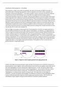
One use of light microscopes is measuring the size of microorganisms. To do this, an eyepiece graticule (a
disc with 100 equal divisions of no set scale) is placed in the eyepiece. To calibrate it, it is aligned with the
scale on a stage micrometre and the length of the micrometre’s scale (for example 1000um) divided by the
number of divisions equal to it; the equation for this is ‘one graticule division = number of micrometres /
number of graticule divisions’ it is also known as the ‘magnification factor’. This must be repeated after each
change in magnification. (7) To calculate the actual size of a specimen, compare the area to be measured to
the eyepiece graticule, view how many divisions is equal to the specimen’s length, then multiply this by the
magnification factor, or the length of each graticule division. The equation for this is ‘actual size (um) =
graticule divisions x magnification factor’.
Figure 1. Diagram to show eyepiece graticule and stage graticule. (8)
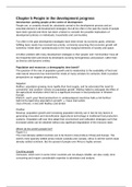
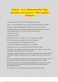
Another useful light microscopy technique is gram staining. This procedure aims to identify a bacterium to
be gram positive or gram negative. Gram positive bacteria retain the first stain applied, crystal violet, due to
their thick peptidoglycan cell wall, whereas gram negative bacteria – as their cell walls are high in lipids
(lipopolysaccharide layer) and have only thin layers of peptidoglycan - can be washed of the stain and are
instead stained red by the safranin counterstain. This difference is particularly relevant in medicine.
Peptidoglycan is a substance that is specific to bacterial cells – this makes them an ideal target for
antibiotics, as animal cells should not be damaged. The difference in thickness of their peptidoglycan layers
results in antibiotics generally being more effective against gram positive bacteria. (10) Therefore, the
identification of gram negative and gram positive bacteria is essential in medical treatment.
, Figure 2. Diagram to show gram negative and gram positive bacteria structure. (9)
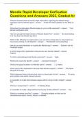
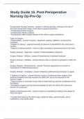
Light microscopes can be used to visualise fungal specimens also. One method of which involves pressing a
piece of scotch tape attached to a cocktail stick onto the mycelial surface in order to collect a sample of the
fungus. This is then attached to a slide, stained with the cotton blue stain and covered with a cover glass.
When viewing these slides under the light microscope, several structures can be viewed; many hyphae,
which look like long, hair-like strands, can often be seen, small spots called spores can also be seen, and
sometimes sporangia, spore-producing structures, are seen as well. Hyphae can be identified as ‘septate’ or
‘aseptate’, depending on whether they are divided into sections by regularly occurring cross-walls (septa).
Generally, this procedure can be used in identification of pathogenic fungi for medical reasons, as to find a
favourable method of treatment. Some fungal infections, for example zygomycete, are very characteristic in
appearance.
Figure 3. Fungi sample under a light microscope
There are two types of electron microscopes – transmission electron microscopes (TEM) and scanning
electron microscopes (SEM). TEMs work by using electromagnetic lenses to force electrons into thin beams,
directed through a specimen within a vacuum. The un-scattered electrons form a ‘shadow image’ of the
specimen. (12) SEMs follow the same principle, but instead detectors collect the x-rays, primary backscatter
electrons, secondary electrons and auger electrons which are emitted when the beam of electrons meets
the specimen - these processes are further explained later. SEMs have large depths of fields, meaning more
of the specimen can be clearly viewed at once. Unlike TEMs, SEMs use electromagnets rather than lenses,
providing more control over the magnification. TEMS can magnify higher than any other microscope, as they
have a much higher resolution. (13) SEMs also have their advantages, they can produce highly detailed
, three-dimensional images and show superficial structures of compounds and elements, as well as shape, size
and surface features of specimens. (14)
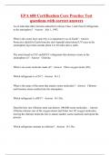
Figure 4. Diagram to show the three domains of life ‘phylogenetic tree of life’
Figure 4 above shows the three-domain system of classification, which splits all living organisms into three
categories; bacteria – single-celled prokaryotic (do not have nuclei) organisms with no membrane-bound
organelles -, archaea – similar to bacteria but differing in their RNA and cell walls – and eukaryota -
unicellular or multicellular organisms containing membrane bound nuclei and organelles. (15) These groups
are further divided into the 5 kingdoms: animalia, plantae, fungi, protoctista, prokaryotae. (16)
Bacteria are microscopic, single-celled organisms with no membrane-bound organelles. They contain a cell
wall made of peptidoglycan sacculus, a feature unique to bacterial cells. The exact composition of this differs
between species but it is generally comprised of glycan strands held together by peptides. (17) They lack
membrane-bound organelles but have ribosomes, cytoplasm and a plasma membrane in common with
eukaryotic cells. (18) DNA is loose in the cell, but extra, beneficial genes are often stored in loops called
plasmids. A structure found in many bacteria species is the capsule – an outside layer comprised of
polysaccharide chains which usually serves to protect the cell from host-cell defence mechanisms. (19) The
general structure and morphology of bacteria is used as the primary method of classification of bacteria. The
three main shapes are the cocci (spherical shape), bacilli (rod shapes) and spirals. As seen in figure 7 below,
each shape exists in various forms, for example cocci can exist alone, in pairs, fours, cubes, groups and
chains. The presence of specialised structures such as flagella (for movement) and pili (for surface adhesion)
can also be used to characterise them; flagellate rods such as salmonella typhi are a type of bacilli shape
which has flagella. Mode of nutrition is also used to classify bacteria, not only down to autotrophs and
heterotrophs but even specific types of feeding such as phagocytosis, and types of food such as decaying
organic matter (saprotrophs). Autotrophic bacteria use a variety of cycles to produce their own nutrion using
carbon dioxide as a source of carbon. (20) Heterotrophic bacteria often feed on decaying plant or animal
matter and waste products, these are known as decomposers and are essential to all ecosystems. Some feed
off living organisms, known as parasitic (harmful to the host) or symbiotic (beneficial to the host)
Microscopy has a range of uses within microbiology. One type of microscope, the light microscope, is
particularly useful in viewing living organisms, as well as organelles such as the nucleus, cytoplasm, cell
membrane, cell wall and chloroplasts - a stain will be needed in order to view these. (4) They are relatively
inexpensive and require little training to use, as the slides are straight forward to prepare and the
microscopes themselves simple to use. However, they generally magnify no more than x2000, which means
smaller organelles such as ribosomes cannot be observed. Electron microscopes can visualise much smaller
details such as viruses and the structure of protein molecules. They are found almost exclusively in hospitals
and industrial labs as they are very expensive, difficult to operate, and take up a lot of space. Slides are very
difficult to prepare, particularly cell cultures and biological samples, as they are more likely soft and moist,
making them unsuited to the vacuum conditions needed for electron microscopes to work. (6) Also, in the
case of the transmission electron microscopes (TEM) specimens undergo other complex procedures as they
must be ultra-thin in order to be visualised. (5)
One use of light microscopes is measuring the size of microorganisms. To do this, an eyepiece graticule (a
disc with 100 equal divisions of no set scale) is placed in the eyepiece. To calibrate it, it is aligned with the
scale on a stage micrometre and the length of the micrometre’s scale (for example 1000um) divided by the
number of divisions equal to it; the equation for this is ‘one graticule division = number of micrometres /
number of graticule divisions’ it is also known as the ‘magnification factor’. This must be repeated after each
change in magnification. (7) To calculate the actual size of a specimen, compare the area to be measured to
the eyepiece graticule, view how many divisions is equal to the specimen’s length, then multiply this by the
magnification factor, or the length of each graticule division. The equation for this is ‘actual size (um) =
graticule divisions x magnification factor’.
Figure 1. Diagram to show eyepiece graticule and stage graticule. (8)
Another useful light microscopy technique is gram staining. This procedure aims to identify a bacterium to
be gram positive or gram negative. Gram positive bacteria retain the first stain applied, crystal violet, due to
their thick peptidoglycan cell wall, whereas gram negative bacteria – as their cell walls are high in lipids
(lipopolysaccharide layer) and have only thin layers of peptidoglycan - can be washed of the stain and are
instead stained red by the safranin counterstain. This difference is particularly relevant in medicine.
Peptidoglycan is a substance that is specific to bacterial cells – this makes them an ideal target for
antibiotics, as animal cells should not be damaged. The difference in thickness of their peptidoglycan layers
results in antibiotics generally being more effective against gram positive bacteria. (10) Therefore, the
identification of gram negative and gram positive bacteria is essential in medical treatment.
, Figure 2. Diagram to show gram negative and gram positive bacteria structure. (9)
Light microscopes can be used to visualise fungal specimens also. One method of which involves pressing a
piece of scotch tape attached to a cocktail stick onto the mycelial surface in order to collect a sample of the
fungus. This is then attached to a slide, stained with the cotton blue stain and covered with a cover glass.
When viewing these slides under the light microscope, several structures can be viewed; many hyphae,
which look like long, hair-like strands, can often be seen, small spots called spores can also be seen, and
sometimes sporangia, spore-producing structures, are seen as well. Hyphae can be identified as ‘septate’ or
‘aseptate’, depending on whether they are divided into sections by regularly occurring cross-walls (septa).
Generally, this procedure can be used in identification of pathogenic fungi for medical reasons, as to find a
favourable method of treatment. Some fungal infections, for example zygomycete, are very characteristic in
appearance.
Figure 3. Fungi sample under a light microscope
There are two types of electron microscopes – transmission electron microscopes (TEM) and scanning
electron microscopes (SEM). TEMs work by using electromagnetic lenses to force electrons into thin beams,
directed through a specimen within a vacuum. The un-scattered electrons form a ‘shadow image’ of the
specimen. (12) SEMs follow the same principle, but instead detectors collect the x-rays, primary backscatter
electrons, secondary electrons and auger electrons which are emitted when the beam of electrons meets
the specimen - these processes are further explained later. SEMs have large depths of fields, meaning more
of the specimen can be clearly viewed at once. Unlike TEMs, SEMs use electromagnets rather than lenses,
providing more control over the magnification. TEMS can magnify higher than any other microscope, as they
have a much higher resolution. (13) SEMs also have their advantages, they can produce highly detailed
, three-dimensional images and show superficial structures of compounds and elements, as well as shape, size
and surface features of specimens. (14)
Figure 4. Diagram to show the three domains of life ‘phylogenetic tree of life’
Figure 4 above shows the three-domain system of classification, which splits all living organisms into three
categories; bacteria – single-celled prokaryotic (do not have nuclei) organisms with no membrane-bound
organelles -, archaea – similar to bacteria but differing in their RNA and cell walls – and eukaryota -
unicellular or multicellular organisms containing membrane bound nuclei and organelles. (15) These groups
are further divided into the 5 kingdoms: animalia, plantae, fungi, protoctista, prokaryotae. (16)
Bacteria are microscopic, single-celled organisms with no membrane-bound organelles. They contain a cell
wall made of peptidoglycan sacculus, a feature unique to bacterial cells. The exact composition of this differs
between species but it is generally comprised of glycan strands held together by peptides. (17) They lack
membrane-bound organelles but have ribosomes, cytoplasm and a plasma membrane in common with
eukaryotic cells. (18) DNA is loose in the cell, but extra, beneficial genes are often stored in loops called
plasmids. A structure found in many bacteria species is the capsule – an outside layer comprised of
polysaccharide chains which usually serves to protect the cell from host-cell defence mechanisms. (19) The
general structure and morphology of bacteria is used as the primary method of classification of bacteria. The
three main shapes are the cocci (spherical shape), bacilli (rod shapes) and spirals. As seen in figure 7 below,
each shape exists in various forms, for example cocci can exist alone, in pairs, fours, cubes, groups and
chains. The presence of specialised structures such as flagella (for movement) and pili (for surface adhesion)
can also be used to characterise them; flagellate rods such as salmonella typhi are a type of bacilli shape
which has flagella. Mode of nutrition is also used to classify bacteria, not only down to autotrophs and
heterotrophs but even specific types of feeding such as phagocytosis, and types of food such as decaying
organic matter (saprotrophs). Autotrophic bacteria use a variety of cycles to produce their own nutrion using
carbon dioxide as a source of carbon. (20) Heterotrophic bacteria often feed on decaying plant or animal
matter and waste products, these are known as decomposers and are essential to all ecosystems. Some feed
off living organisms, known as parasitic (harmful to the host) or symbiotic (beneficial to the host)