LECTURE 21: TOOLS IN TUMOR IMMUNOLOGY/SINGLE CELL TECH.
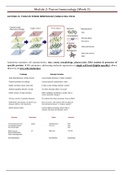
Cytometry examines cell characteristics: size, count, morphology, phase/cycle, DNA content & presence of
specific protein HD cytometry: addressing molecule expression at single cell level (highly specific); allows
discovery of rare cells/molecules
, Module 2: Tumor Immunology (Week 3)
Cytometry allows examination of cells as
single cell/individual: behavioral change
upon certain stimulation (e.g. oncogenic
mutation) & response of each cell to certain
drug
Fundamental principle of cytometry (conventional)
Determination of normal & abnormal
functions of the cells
Single cell suspension goes through the
sheath laser dispersion (light source)
collected by optics system (prism, mirror,
filter) electronic amplification
analysis
Examining the morphology & functional
characteristics from tissue biopsy
Cells extracted from tissue sample is loaded with
antigen put through the suspension tube
analysis of morphology & function at the same time
(TOF-MS)
Different wavelength/light source to examine
different function composite/concomitant
analysis (e.g. cell status, immune cell types, regulatory
functions/Treg, IDO, etc, stromal conditions)
, Module 2: Tumor Immunology (Week 3)
HD Cytometry (less noise d/t reduced scatter)
Metal-elements based: mass cytometry (using rare natural elements)
Signal detection affected by: antigen level, sensitivity of sensor, background signals & cross-talk between
abundance sensitivity, isotope purity, and metal ion oxidation
, Module 2: Tumor Immunology (Week 3)
Fluorescence-based:
BD Symphony (each light source emits 1 wavelength): risk of cross-talk & spillover >>> (non-specific)
using PMT (photon multiplier tube)
PMT


