The Trigeminal Nerve (CN V)
The trigeminal nerve is the largest of the cranial nerves
It has both sensory and motor components, and is the main sensory nerve for
the head
- Sensory: the three terminal branches of the trigeminal nerve innervate the
skin, mucous membranes and sinuses of the face. Their distribution is
similar to the dermatome supply from the spinal nerves.
- Motor: the only branch of the trigeminal nerve to have motor fibres is the
mandibular branch. It acts to innervate the muscles of mastication,
medial and lateral pterygoid, masseter and temporalis muscles. It
also supplies derivatives from the 1st pharyngeal arch: the anterior belly
of digastric, tensor veli palatini and the tensor tympani.
Anatomical course:
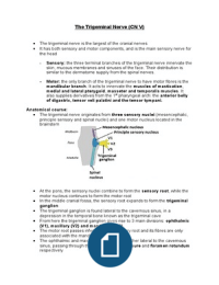
The trigeminal nerve originates from three sensory nuclei (mesencephalic,
principle sensory and spinal nuclei) and one motor nucleus located in the
brainstem
At the pons, the sensory nuclei combine to form the sensory root, while the
motor nucleus continues to form the motor root
In the middle cranial fossa, the sensory root expands to form the trigeminal
ganglion
The trigeminal ganglion is found lateral to the cavernous sinus, in a
depression in the temporal bone known as the trigeminal cave
From here the trigeminal ganglion gives rise to 3 main divisions: ophthalmic
(V1), maxillary (V2) and mandibular (V3)
The motor root passes inferiorly to the sensory root and its fibres are only
associated with the mandibular division (V2)
The ophthalmic and maxillary nerve travel together lateral to the cavernous
sinus, passing through the superior orbital fissure and foramen rotundum
respectively
The trigeminal nerve is the largest of the cranial nerves
It has both sensory and motor components, and is the main sensory nerve for
the head
- Sensory: the three terminal branches of the trigeminal nerve innervate the
skin, mucous membranes and sinuses of the face. Their distribution is
similar to the dermatome supply from the spinal nerves.
- Motor: the only branch of the trigeminal nerve to have motor fibres is the
mandibular branch. It acts to innervate the muscles of mastication,
medial and lateral pterygoid, masseter and temporalis muscles. It
also supplies derivatives from the 1st pharyngeal arch: the anterior belly
of digastric, tensor veli palatini and the tensor tympani.
Anatomical course:
The trigeminal nerve originates from three sensory nuclei (mesencephalic,
principle sensory and spinal nuclei) and one motor nucleus located in the
brainstem
At the pons, the sensory nuclei combine to form the sensory root, while the
motor nucleus continues to form the motor root
In the middle cranial fossa, the sensory root expands to form the trigeminal
ganglion
The trigeminal ganglion is found lateral to the cavernous sinus, in a
depression in the temporal bone known as the trigeminal cave
From here the trigeminal ganglion gives rise to 3 main divisions: ophthalmic
(V1), maxillary (V2) and mandibular (V3)
The motor root passes inferiorly to the sensory root and its fibres are only
associated with the mandibular division (V2)
The ophthalmic and maxillary nerve travel together lateral to the cavernous
sinus, passing through the superior orbital fissure and foramen rotundum
respectively