Varicose Vein Examination
General inspection:
Expose the patient. You should ask to remove their trousers to perform a
lower limb examination. Also offer a chaperone.
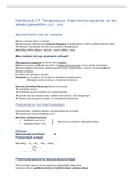
Varicose veins usually follow the path of 2 major veins in the leg:
1. Long saphenous vein
2. Short saphenous vein
Lipodermatosclerosis – areas of brown/red/orange thickened skin cause by
fibrosis of the subcutaneous fat.
Saphena varix – dilation of the to of the long saphenous vein due to valvular
incompetence.
Venous eczema – caused by leakage of venous content which is irritant to
skin.
Venous flare – venous tortuosities around the malleolus.
Skin pigmentation – caused by haemosiderin deposition in the lower limbs.
Atrophie blanche – white scar tissue with dotted capillaries.
Odema
Venous ulcers
Palpation:
Feel for temperature disparity and tenderness (heat and tenderness are signs
of superficial thrombophlebitis).
Pitting odema.
General inspection:
Expose the patient. You should ask to remove their trousers to perform a
lower limb examination. Also offer a chaperone.
Varicose veins usually follow the path of 2 major veins in the leg:
1. Long saphenous vein
2. Short saphenous vein
Lipodermatosclerosis – areas of brown/red/orange thickened skin cause by
fibrosis of the subcutaneous fat.
Saphena varix – dilation of the to of the long saphenous vein due to valvular
incompetence.
Venous eczema – caused by leakage of venous content which is irritant to
skin.
Venous flare – venous tortuosities around the malleolus.
Skin pigmentation – caused by haemosiderin deposition in the lower limbs.
Atrophie blanche – white scar tissue with dotted capillaries.
Odema
Venous ulcers
Palpation:
Feel for temperature disparity and tenderness (heat and tenderness are signs
of superficial thrombophlebitis).
Pitting odema.