View metadata, citation and similar papers at core.ac.uk brought to you by CORE
provided by Southeast Asian Fisheries Development Center, Aquaculture Department Institutional.
Southeast Asian Fisheries Development Center Aquaculture Department
SEAFDEC/AQD Institutional Repository http://repository.seafdec.org.ph
Books Health Management in Aquaculture
2001
Bacterial diseases
Alapide-Tendencia, Eleonor V.
Aquaculture Department, Southeast Asian Fisheries Development Center
Alapide-Tendencia, E. V., & de la Peña, L. D. (2001). Bacterial diseases. In G. D. Lio-Po, C. R.
Lavilla, & E. R. Cruz-Lacierda (Eds.), Health management in aquaculture (pp. 25-41). Tigbauan,
Iloilo, Philippines: Aquaculture Department, Southeast Asian Fisheries Development Center.
http://hdl.handle.net/10862/726
Downloaded from http://repository.seafdec.org.ph, SEAFDEC/AQD's Institutional Repository
,○ ○ ○ ○ ○ ○ ○ ○ ○ ○ ○ ○ ○ ○ ○ ○ ○ ○ ○ ○ ○ ○ ○ ○ ○ ○ ○ ○ ○ ○ ○ ○ ○ ○ ○ ○ ○ ○ ○ ○ ○ ○ ○ ○ ○ ○ ○ ○ ○ ○ ○ ○ ○ ○ ○ ○ ○ ○ ○ ○ ○
3
CHAPTER THREE Bacterial diseases
Eleonor V. Alapide-Tendencia and Leobert D. de la Peña
Diseases caused by bacteria may cause heavy mortality in both wild and cul-
tured fish and crustaceans. Bacteria are found everywhere in the aquatic envi-
ronment. Most bacterial disease agents are part of the normal microflora of the
marine environment and are generally considered as secondary or opportunis-
tic pathogens. Almost all fish bacterial pathogens are capable of independent
existence outside the fish. There are only a few obligatory pathogens. Even
these, however, are capable of living for a long time in the tissues of their host
without causing injury. Clinical infections and disease usually occur only after
the onset of some major changes in the physiology or body of the host. Thus, to
understand bacterial diseases of fish, one must understand the relationship of
bacteria with their host and with their environment.
As in all animal production systems, bacterial disease is one of the major prob-
lems facing production, development and expansion of the aquaculture indus-
try. The control of disease is particularly difficult because fish are often farmed
in systems where production is dependent on natural environmental condi-
tions. Changes or deterioration in the aquatic environment cause most of the
bacterial diseases encountered, and environmental effects give rise to many
other adverse conditions. A second major constraint on disease control is the
relatively limited number of therapeutic agents available for the control of bac-
terial disease agents. Even recommended therapies and preventive measures
pose limitations. Their application to aquatic animals is often difficult in actual
practice, and sometimes impossible to implement.
Outbreaks of major bacterial diseases in aquaculture can be significantly re-
duced if proper attention is paid to good husbandry practices and the mainte-
nance of optimum environmental conditions, especially water quality. Another
important consideration involves identifying the predisposing factors that may
lead to a disease state. Once predisposing factors are identified, appropriate
corrective measures should be initiated in the culture system.
Most bacterial disease show similar signs, especially in fishes. Bacterial infec-
tion may appear on the skin or fins of fish, exoskeleton or appendages of crus-
taceans, in the muscles and in the internal organs. In nearly all cases, red spots,
brown or black spots, or necrotic tissues can be observed. Inflammation may
also occur. Proper identification of the causative agent is important to ensure
successful treatment.
, WHAT ARE B
WHAT BAA CTERIA?
Bacteria are not visible to the naked eye. These microorganisms are of very
small dimensions, usually between 0.5 and 10 microns (µm). But, when bacte-
a b
ria multiply in great numbers on a solid medium, they form visible colonies
representing millions or billions of individual cells. The cells can be seen only
under a microscope from a smear stained with a dye on a microscope slide.
Bacteria differ from other cells in that they are prokaryotic (lacking a nuclear
c membrane). The nucleus occupies the center of the cell. All its genetic material
is linked in a single chromosome. The cytoplasm is densely packed with RNA
and is finely granular because of the presence of ribosome. The nucleus/ cyto-
plasm complex is packaged in a complex envelope or integument. Its inner-
most layer is the thin cytoplasmic membrane (plasmalemma). Outside the
membrane is a rigid cell wall. Some bacterial pathogens develop a capsule out-
d e side of the cell wall, which is usually associated with the virulence or infective
ability of the organism. Many of the pathogenic bacteria are flagellated and a
few have no flagella for locomotion. Some move by body flexing or gliding.
Some bacteria produce enzymes called extracellular products or ECP, which
are associated with the microorganism’s virulence. Extracellular products are
highly toxic to fish and crustaceans and they contain proteases, hemolysins,
exohemagglutinins and cytotoxins.
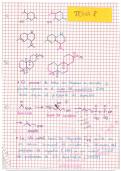
Figure 3-1. The different shapes and ar-
rangements of bacteria: (a) cocci in Bacteria reproduce asexually by binary fission. That is, they multiply by an
streptococcal arrangement, (b) cocci in elongation of the cell followed by a division.
staphylococcal arrangement, (c) bacilli,
(d) several kinds of spirilla, and (e) The most common method used to detect the presence of bacteria is by gram
comma-shaped vibrios stain. The gram stain classifies bacteria into two groups: the gram positive and
the gram negative. Gram-positive bacteria are those that possess a thick pepti-
doglycan cell wall which will retain the initial crystal violet stain during wash-
ing with 95% alcohol. Gram-negative bacteria are those that possess a uni-
molecular peptidoglycan cell wall bounded on one side by the cytoplasmic
membrane and on the other side by the outer membrane; such cells are decol-
orized by 95% alcohol and take up the secondary stain. To identify bacterium,
a pure culture should be obtained, containing a single species and not a mix-
ture of different kinds of bacteria.
In classifying bacteria, one needs to pay attention to the cell shape. There are
three distinct cell forms: cocci, baccili and spiral (Fig.3-1). Cocci are spherical
cells and exist in several patterns or groupings which are specific to the genus.
Paired cocci are called diplococcus, while those in long chains are called strep-
tococcus. Irregularly grouped cocci are called staphylococcus. Longer and cy-
lindrical bacteria are known as bacilli or rods. Cells that are between the coc-
cus and the bacillus in shape are called coccobacilli. The short, curved rods are
the vibrios. When more than one curvature is observed, it is called spirilla.
Most bacteria that cause disease in fish and crustaceans are rod-shaped. Figure
3-1 shows the different shapes and arrangements of bacteria.
The shape, size and color of a given colony are also important in identification.
The bacterial colony surface texture, whether rough, smooth or mucoid, should
26 Health Management in Aquaculture
provided by Southeast Asian Fisheries Development Center, Aquaculture Department Institutional.
Southeast Asian Fisheries Development Center Aquaculture Department
SEAFDEC/AQD Institutional Repository http://repository.seafdec.org.ph
Books Health Management in Aquaculture
2001
Bacterial diseases
Alapide-Tendencia, Eleonor V.
Aquaculture Department, Southeast Asian Fisheries Development Center
Alapide-Tendencia, E. V., & de la Peña, L. D. (2001). Bacterial diseases. In G. D. Lio-Po, C. R.
Lavilla, & E. R. Cruz-Lacierda (Eds.), Health management in aquaculture (pp. 25-41). Tigbauan,
Iloilo, Philippines: Aquaculture Department, Southeast Asian Fisheries Development Center.
http://hdl.handle.net/10862/726
Downloaded from http://repository.seafdec.org.ph, SEAFDEC/AQD's Institutional Repository
,○ ○ ○ ○ ○ ○ ○ ○ ○ ○ ○ ○ ○ ○ ○ ○ ○ ○ ○ ○ ○ ○ ○ ○ ○ ○ ○ ○ ○ ○ ○ ○ ○ ○ ○ ○ ○ ○ ○ ○ ○ ○ ○ ○ ○ ○ ○ ○ ○ ○ ○ ○ ○ ○ ○ ○ ○ ○ ○ ○ ○
3
CHAPTER THREE Bacterial diseases
Eleonor V. Alapide-Tendencia and Leobert D. de la Peña
Diseases caused by bacteria may cause heavy mortality in both wild and cul-
tured fish and crustaceans. Bacteria are found everywhere in the aquatic envi-
ronment. Most bacterial disease agents are part of the normal microflora of the
marine environment and are generally considered as secondary or opportunis-
tic pathogens. Almost all fish bacterial pathogens are capable of independent
existence outside the fish. There are only a few obligatory pathogens. Even
these, however, are capable of living for a long time in the tissues of their host
without causing injury. Clinical infections and disease usually occur only after
the onset of some major changes in the physiology or body of the host. Thus, to
understand bacterial diseases of fish, one must understand the relationship of
bacteria with their host and with their environment.
As in all animal production systems, bacterial disease is one of the major prob-
lems facing production, development and expansion of the aquaculture indus-
try. The control of disease is particularly difficult because fish are often farmed
in systems where production is dependent on natural environmental condi-
tions. Changes or deterioration in the aquatic environment cause most of the
bacterial diseases encountered, and environmental effects give rise to many
other adverse conditions. A second major constraint on disease control is the
relatively limited number of therapeutic agents available for the control of bac-
terial disease agents. Even recommended therapies and preventive measures
pose limitations. Their application to aquatic animals is often difficult in actual
practice, and sometimes impossible to implement.
Outbreaks of major bacterial diseases in aquaculture can be significantly re-
duced if proper attention is paid to good husbandry practices and the mainte-
nance of optimum environmental conditions, especially water quality. Another
important consideration involves identifying the predisposing factors that may
lead to a disease state. Once predisposing factors are identified, appropriate
corrective measures should be initiated in the culture system.
Most bacterial disease show similar signs, especially in fishes. Bacterial infec-
tion may appear on the skin or fins of fish, exoskeleton or appendages of crus-
taceans, in the muscles and in the internal organs. In nearly all cases, red spots,
brown or black spots, or necrotic tissues can be observed. Inflammation may
also occur. Proper identification of the causative agent is important to ensure
successful treatment.
, WHAT ARE B
WHAT BAA CTERIA?
Bacteria are not visible to the naked eye. These microorganisms are of very
small dimensions, usually between 0.5 and 10 microns (µm). But, when bacte-
a b
ria multiply in great numbers on a solid medium, they form visible colonies
representing millions or billions of individual cells. The cells can be seen only
under a microscope from a smear stained with a dye on a microscope slide.
Bacteria differ from other cells in that they are prokaryotic (lacking a nuclear
c membrane). The nucleus occupies the center of the cell. All its genetic material
is linked in a single chromosome. The cytoplasm is densely packed with RNA
and is finely granular because of the presence of ribosome. The nucleus/ cyto-
plasm complex is packaged in a complex envelope or integument. Its inner-
most layer is the thin cytoplasmic membrane (plasmalemma). Outside the
membrane is a rigid cell wall. Some bacterial pathogens develop a capsule out-
d e side of the cell wall, which is usually associated with the virulence or infective
ability of the organism. Many of the pathogenic bacteria are flagellated and a
few have no flagella for locomotion. Some move by body flexing or gliding.
Some bacteria produce enzymes called extracellular products or ECP, which
are associated with the microorganism’s virulence. Extracellular products are
highly toxic to fish and crustaceans and they contain proteases, hemolysins,
exohemagglutinins and cytotoxins.
Figure 3-1. The different shapes and ar-
rangements of bacteria: (a) cocci in Bacteria reproduce asexually by binary fission. That is, they multiply by an
streptococcal arrangement, (b) cocci in elongation of the cell followed by a division.
staphylococcal arrangement, (c) bacilli,
(d) several kinds of spirilla, and (e) The most common method used to detect the presence of bacteria is by gram
comma-shaped vibrios stain. The gram stain classifies bacteria into two groups: the gram positive and
the gram negative. Gram-positive bacteria are those that possess a thick pepti-
doglycan cell wall which will retain the initial crystal violet stain during wash-
ing with 95% alcohol. Gram-negative bacteria are those that possess a uni-
molecular peptidoglycan cell wall bounded on one side by the cytoplasmic
membrane and on the other side by the outer membrane; such cells are decol-
orized by 95% alcohol and take up the secondary stain. To identify bacterium,
a pure culture should be obtained, containing a single species and not a mix-
ture of different kinds of bacteria.
In classifying bacteria, one needs to pay attention to the cell shape. There are
three distinct cell forms: cocci, baccili and spiral (Fig.3-1). Cocci are spherical
cells and exist in several patterns or groupings which are specific to the genus.
Paired cocci are called diplococcus, while those in long chains are called strep-
tococcus. Irregularly grouped cocci are called staphylococcus. Longer and cy-
lindrical bacteria are known as bacilli or rods. Cells that are between the coc-
cus and the bacillus in shape are called coccobacilli. The short, curved rods are
the vibrios. When more than one curvature is observed, it is called spirilla.
Most bacteria that cause disease in fish and crustaceans are rod-shaped. Figure
3-1 shows the different shapes and arrangements of bacteria.
The shape, size and color of a given colony are also important in identification.
The bacterial colony surface texture, whether rough, smooth or mucoid, should
26 Health Management in Aquaculture