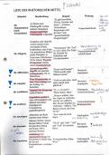
Management of Patient with
Burns
, Review of Anatomy of the Skin
• The skin is composed of three layers: epidermis, dermis, and
subcutaneous tissue
Epidermis
outermost layer of stratified epithelial cells, composed predomin
keratinocytes. It ranges in thickness from about 0.1 mm on the ey
about 1 mm on the palms of the hands and soles of the feet.
Four distinct
• layers compose the epidermis; from innermost to outermost, they
stratum germinativum, stratum granulosum, stratum lucidum, and s
corneum.
Melanocytes are the special cells of the epidermis that are primar
involved in producing the pigment melanin, which colors the skin.
Production of melanin is controlled by a hormone secreted from th
hypothalamus of the brain called melanocyte-stimulating hormone.
,
Two other types of cells in the epidermis: Merkel and Langerh
cells. Merkel cells are receptors that transmit stimuli to the axon
through a chemical synapse. Langerhans cells - play a significa
in cutaneous immune system reactions.
Epidermis is thickest over the palms of the hands and soles o
feet and contains increased amounts of keratin. The thickness
epidermis can increase with use and can result in calluses form
the hands or corns forming on the feet.
The junction of the epidermis and dermis is an area of many
undulations and furrows called rete ridges. This junction anchors
the free exchange of essent
epidermis to the dermis and permits
nutrients between the two layers.
,
Dermis:
The dermis makes up the largest portion of the skin, providing strength an
structure. It is composed of two layers: papillary and reticular. The papi
dermis lies directly beneath the epidermis and is composed primarily of fi
cells capable of producing one form of collagen, a component of connect
tissue. The reticular layer lies beneath the papillary layer and also produc
collagen and elastic bundles. The dermis also contains blood and lymph
nerves, sweat and sebaceous glands, and hair roots. The dermis is often
to as the “true skin.”
Subcutaneous Tissue (hypodermis):
innermost layer of the skin.
It is primarily adipose tissue, which provides a cushion between the skin layers
muscles, and bones. It promotes skin mobility, molds body contours, and insula
body.
Fat is deposited and distributed according to the person’s
gender and in part acco
for the difference in body shape between men and women
The subcutaneous tissues and the amount of fat deposited are important factors in
body temperature regulation