100% tevredenheidsgarantie Direct beschikbaar na betaling Zowel online als in PDF Je zit nergens aan vast
logo-home
Samenvatting Anatomie 4 €10,49
In winkelwagen

Samenvatting

Samenvatting Anatomie 4

 71 keer bekeken  5 keer verkocht

Samenvatting anatomie 4 dissecties van alle leerpaden (in het Engels). Dit document bevat afbeeldingen uit de demovideo's, Acland anatomy video's en overige nuttige afbeeldingen ter verduidelijking. Alle termen die gekend moeten zijn voor het examen, worden besproken.

Voorbeeld 10 van de 159  pagina's

  • 17 juni 2022
  • 159
  • 2021/2022
  • Samenvatting
Alle documenten voor dit vak (21)
avatar-seller
JBtns
ANATOMY 4: DISSECTIONS
Upper extremity
1 The shoulder (cingulum pectorale)
1.1 Bones of the shoulder
https://aclandanatomy.com/MultimediaPlayer.aspx?multimediaid=10528033

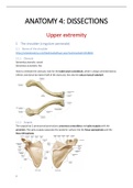
1.1.1 Clavicula
Extremitas sternalis: round
Extremitas acromialis: flat

How to orientate the clavicula: look for the tuberculum conoideum, which is always oriented lateral,
inferior and dorsal (on lateral half of the clavicula). See also the sulcus musculi subclavii.


1.1.2 Scapula
The scapula has 2 pronounced protrusions: processus coracoideus and spina scapula with the
acromion. The spina scapula separates the posterior surface into the fossa supraspinata and the
fossa infraspinata.


1

,The cavitas glenoidalis is the articular surface for the shoulder joint. Above the surface, the
tuberculum supraglenoidale and below the tuberculum infraglenoidale can be found. On these
protrusions, muscles will attach. The cavitas glenoidalis is very shallow, therefore it is lined by the
labrum glenoidale to deepen the socket.

1.1.3 Humerus
The humerus consists of multiple parts:

- Caput humeri: facing medial
- Collum anatomicum
- Tuberculum majus: positioned laterally and has 3 facets (upper,
medial and lower) for the insertion of 3 muscles (m.
supraspinatus, m. infrspinatus and m. teres minor)
- Tuberculum minus: faces ventrally
- Sulcus intertubercularis: this is the groove between the 2
tubercles. The tendon of the caput longum of the m. biceps
brachii runs through it.
- Corpus humeri (diaphysis) with the tuberositas deltoidea,
which is a V-shaped tuberosity halfway the corpus on the
lateral side. Posterior from this, the sulcus nervi radialis can be
found.
- Condylus humeri: the epiphysis here is formed out of 2 joint
surfaces: trochlea humeri (medial) and capitulum humeri
(lateral), and 2 epicondyli (medialis and lateralis). Medial of
the trochlea humeri, there is the sulcus n. ulnaris.


1.2 Joints of the shoulder
https://aclandanatomy.com/MultimediaPlayer.aspx?multimediaId=10528039

Between the shoulder girdle and the trunk, there are 2 synovial joints:

- Sternoclavicular joint: with lig. Sternoclaviculare (anterius and posterius) and lig.
Costoclaviculare.
- Acromioclavicular joint: with lig. Coracoacromiale, lig. Acromioclaviculare and the ligg.
Coracoclaviculare, which consists of lig. Trapezoideum and lig. Conoideum.


2

,The capsula articularis of the shoulder joint attaches outside the labrum glenoidale. This tissue
surrounds the art. Glenohumeralis. It is strengthened by the lig. Coracohumerale and ligg.
Glenohumerale (superius, medium and inferius).


1.3 Muscles
The muscles connecting the trunk to the shoulder girdle are subdivided into 2 functional groups:

- Trunk-shoulder girdle muscles which have their origo onto the trunk and insertion on the
scapula and clavicula
- The trunk-upper arm muscles with their origin onto the trunk and their insertion on the
humerus

1.3.1 Muscles from scapula to humerus
https://aclandanatomy.com/MultimediaPlayer.aspx?multimediaid=10528045

Very deep, below anything else, lie the long tendon of the m. biceps and triceps brachii. The biceps
tendon goes through the capsula and attaches at the tuberculum supraglenoidalis, and the triceps
tendon at the tuberculum infraglenoidalis.


Rotator cuff:
These muscles stabilize the shoulder joint: (see 3D model)


3

, - M. subscapularis

Origo: facies costalis scapulae

Insertion: tub. minus


→ ventral side of the scapula
- M. supraspinatus
Origo: fossa supraspinata

Insertion: tub. Majus (upper facet)


→ dorsal side
- M. infraspinatus Origo: fossa infraspinata
See next picture
Insertion: tub. Majus (medial facet)


- M. teres minor

Origo: margo lateralis scapulae

Insertion: tub. Majus (lower facet)


Other muscles:
- M. teres major:
Origo: near angulus inferior scapulae

Insertion: crista tub. minoris


- M. coracobrachialis:
Origo: proc. coracoideus

Insertion: tuberositas
coracobrachialis (= medial side
halfway on the humerus)


- M. deltoideus: see infra


4

,1.3.2 Muscles from scapula to trunk
https://aclandanatomy.com/MultimediaPlayer.aspx?multimediaid=10528049

- M. serratus anterior:
Origo: ventral end of rib 2-8

Insertion: ventral side margo
medialis scapulae


- M. trapezius:

Origo: linea nuchae superior up to
protuberantia occipitalis externa +
linea nuchae + proc. spinosi T1-T12

Insertion: lateral part clavicula +
medial border acromion + spina
scapula

- M. levator scapulae: Origo: tub. dorsale proc.
= number 1 on next picture transversarii C1-C4

Insertion: angulus superior
scapulae + margo medialis until
trigonum
- M. rhomboideus minor (2) and major (3):

Origo: proc. spinosi C6-T5
1
2
Insertion: margo medialis
→ minor at the level of spina
3
scapula, major is below that


- M. pectoralis minor:
Origo: ventral part of rib 2-4

Insertion: proc. coracoideus


5

, - M. subclavius:

Origo: border 1st rib and cartilage

Insertion: sulcus subclavius (= at
caudal side clavicula)


- M. omohyoideus:
Origo: margo superior scapulae

Insertion: lateral 1/3 corpus ossis
hyoidei (bottom part)


1.3.3 Large shoulder muscles
https://aclandanatomy.com/MultimediaPlayer.aspx?multimediaid=10528041

- M. pectoralis major

Origo: medial 1/3 clavicula + front
sternum + rib cartilages 1-6

Insertion: anterior edge crista tub.
minoris


- M. latissimus dorsi
Origo: T7-sacrum + posterior crista
iliaca + rib 9-12 (+angulus inferior
scapulae)

Insertion: crista tub. Minoris →
spirals around teres major


- M. deltoideus
Origo: lateral border clavicle + lateral
border acromion + lower border spina
scapula

Insertion: tuberositas deltoidea


→ it has 3 parts that can be distinguished easily. It goes around the whole shoulder.

6

,1.3.4 Arrangement of the thoracic aperture
https://aclandanatomy.com/MultimediaPlayer.aspx?multimediaId=10528047

The main bundle of vessels and nerves lies behind the clavicle and behind both pectoralis muscles.

The superior thoracic aperture is formed by the clavicula, both of the 1st ribs and some vertebrae (C6-
T1). The main spinal nerves in this area are C5-T1


The a. subclavia crosses first rib a bit lateral between first rib and clavicle. The v. subclavia is more to
the middle, right behind the medial end of the clavicle.

The mm. scaleni have an important role as well.


→ the v. subclavia runs in front of the anterior muscle, the a. subclavia behind it.
→ the white things you see on the left picture, are the roots of the brachial plexus that emerge
between the muscles.

Plexus brachialis:
- 3 trunci: superior, medius and inferior
- 3 divisiones: superior, medius and inferior
- 3 cords: fasiculi medialis, lateralis and posterior
- The peripheral nerves: n. medianus, n. axillaris, n. radialis, n. ulnaris and n. musculocutaneus

1.4 Veins
Most major veins run parallel with the majority of the arm arteries. The v. cephalica is an exception.

https://aclandanatomy.com/MultimediaPlayer.aspx?multimediaid=10528055

Vena cephalica:
This vein runs along the anterolateral surface of the m. biceps humeri. It communicates with the v.
basilica via the v. mediana cubiti in the elbow. From there, it continues upward through the sulcus
deltopectoralis (= Mohrenheim-groove). The vein ends in the v. axillaris to form the v. subclavia and
the v. brachiocephalica.

This vein is often used for cannulas and pacemakers etc.


7

,Vena subclavia
The v. subclavia comes up from the arm and will pass underneath the m. pectoralis minor and then
goes over the first rib and under the clavicula and m. subclavius. This vein will form the v.
brachiocephalica by coming together with the v. jugularis interna at the medial border of the 1st rib.


clavicle, m. subclavius and SCM are removed

Vena brachiocephalica:
This vein is formed by the v. jugularis interna and v. subclavia coming together. The 2 brachiocephalic
veins will form the v. cava superior together, a little to the right from the midline.


Part of the chest wall is removed


8

,1.5 Arteries
https://aclandanatomy.com/MultimediaPlayer.aspx?multimediaid=10528053

Brachiocephalic trunk (see trunk):
This structure runs from left to right, and crosses the trachea very high. It arises from the arch of the
aorta.


A. subclavia (see trunk):
It is the first part of an artery, that will proceed as the axillary artery, and further on the brachial
artery. It runs posterior from the M. scalenus anterior, the clavicle and the m. pectoralis minor. The
right a. subclavia originates from the truncus brachiocephalica, together with the a. carotis
communis. On the left side however, it will originate directly from the arch of the aorta. It gives off
several branches.


A. axillaris:
This is the continuation of the a. subclavia (starting from where it passes underneath the clavicula).
It gives of two branches underneath the M. pectoralis minor.


= the two branches, the M. pectoralis is taken away to see.


9

, In the axilla, 3 more branches are formed: a. circumflexa humeri anterior and posterior and the a.
subscapularis.
1: a. subscapularis
2: a. circumflexa humeri posterior
3: a. circumflexa humeri anterior
1
3 2


A. brachialis:
This is the continuation of the a. axillaris and goes down the arm. It is called a. brachialis after it has
passed the inferior border of the m. teres major.

Supply table:


1.6 Plexus brachialis
https://aclandanatomy.com/MultimediaPlayer.aspx?multimediaid=10528059

The plexus provides motoric and sensory innervation of all the arm muscles. It runs through the neck,
armpit and to the arm, but there are 2 exceptions: the m. trapezius is innervated by the n.
accessories and a part of the skin near the armpit is innervated by n. intercostobrachialis.


Right picture: the ventral rami as the nerves emerge. They emerge between the m. scalenus anterior
medius. The upper 2 rami join and the lower 2 rami join. The middle one (C7) stays alone. These 3 big
units are the upper, middle and lower trunk. Each trunk divides in an anterior and posterior


10

Voordelen van het kopen van samenvattingen bij Stuvia op een rij:

√  	Verzekerd van kwaliteit door reviews

√ Verzekerd van kwaliteit door reviews

Stuvia-klanten hebben meer dan 700.000 samenvattingen beoordeeld. Zo weet je zeker dat je de beste documenten koopt!

Snel en makkelijk kopen

Snel en makkelijk kopen

Je betaalt supersnel en eenmalig met iDeal, Bancontact of creditcard voor de samenvatting. Zonder lidmaatschap.

Focus op de essentie

Focus op de essentie

Samenvattingen worden geschreven voor en door anderen. Daarom zijn de samenvattingen altijd betrouwbaar en actueel. Zo kom je snel tot de kern!

Veelgestelde vragen

Wat krijg ik als ik dit document koop?

Je krijgt een PDF, die direct beschikbaar is na je aankoop. Het gekochte document is altijd, overal en oneindig toegankelijk via je profiel.

Tevredenheidsgarantie: hoe werkt dat?

Onze tevredenheidsgarantie zorgt ervoor dat je altijd een studiedocument vindt dat goed bij je past. Je vult een formulier in en onze klantenservice regelt de rest.

Van wie koop ik deze samenvatting?

Stuvia is een marktplaats, je koop dit document dus niet van ons, maar van verkoper JBtns. Stuvia faciliteert de betaling aan de verkoper.

Zit ik meteen vast aan een abonnement?

Nee, je koopt alleen deze samenvatting voor €10,49. Je zit daarna nergens aan vast.

Is Stuvia te vertrouwen?

4,6 sterren op Google & Trustpilot (+1000 reviews)

Afgelopen 30 dagen zijn er 48756 samenvattingen verkocht

Opgericht in 2010, al 15 jaar dé plek om samenvattingen te kopen

Start met verkopen
€10,49  5x  verkocht
  • (0)
In winkelwagen
Toegevoegd