A.P1 A.P2 A.M1 A.M2 A.D1 Criteria's met. This Unit covers all areas of of Musculoskeletal System and the Disorders that are associated with it. Distinction met first time. Highly detailed and informative to obtain a Distinction throughout the whole of Unit 8
`Assignment title; musculoskeletal disorders
Learning aim A
Axial skeleton function and structure
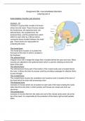
Cranium - [1]
Cranium is a group that consists of 8 bones
which form the head. These 8 bones include
the frontal bone, the sphenoid bone, the
ethmoid bone, the occipital bone, the
temporal bone, and the parietal bone which
attach to each other by a fibrous dense
connective tissue located between the small
gap. These bones are responsible for
protecting the brain.
The frontal bone
The frontal bone function is to protect the
front part of the brain in which is located in
the forehead
The sphenoid bone
Irregular bone with a wedge like shape that is located behind the eyes and nose. Many
muscles are attached to the sphenoid bone which is used for chewing as forms the
base of the skull
The ethmoid bone
The ethmoid bone forms part of the bottom of the cranial cavity and is located behind
the nose. It allows the brain to process smell by providing a passage for olfactory fibers
to pass through
The occipital bone
The occipital bone houses the cerebellum and cerebrum and is located at the back of
the head which the brain with processing vision
The temporal bone
Consists of 2 bones which are at located on each side of the head creating the cavity
sides that forms the brian in which poretcs and houses ear canals and inner ear
structures
The parietal bone
Consists of 2 bones that form the sides and roof of the cranial cavity which sits at the
top of the head. It is responsible for the protection of the brains right and left parietal
lobes
,The mandible bone
The mandible forms the lower jaw and holds the lower teeth in place as well as
assisting in chewing.
The maxilla bones
The maxilla bones are 2 bones that form the upper jaw by connecting the bottom of the
nasal cavity and the upper teeth. Therefore enabling smiling, biting, and chewing.
Vertebral column
The vertebral column is responsible for protecting the spinal cord as well as provides
connection between different muscles and ligaments
Cervical vertebrae - [2]
Cervical vertebrae start at the base of the skull and stretch
down the neck spine. It consists of 7 vertebrae bones
stacked up one another in which are connected to facet
joints.
The cervical vertebrae allows head movements as well as
supports the head's weight.
It enables the head and neck to move backwards,
forwards and tilt from each side by attaching to various
muscles such as trapezius, sternocleidomastoid, and deep
cervical flexors
Thoracic vertebrae - [3]
Thoracic vertebrae consists of 12 thoracic bones in which are found
between the cervical vertebrae and the lumbar vertebrae.
It is responsible for the protection of the spine in which the stacked
vertebrae from the spine create an enclosed and protective canal. It
also supports the chest area by allowing each of the ribs to attach to
each other further protecting important organs such as lungs and
heart along with ribcage. In addition it enables the body to twist and
turn by being made by additional ligaments and facet joints.
,Lumbar vertebrae - [4]
Lumbar vertebrae consists of 5 vertebrae bones stacked upon one another in the lower
back. It sits below the thoracic vertebrae and
above the sacrum bone. They are thick,
large and blocked like bones The lumbar
vertebrae bones are responsible for
protecting the spinal cord by forming a
enclosed canal allowing nerves to pass
through the spinal cord
It supports the majority of the body's weight
and adds stability to the back. It attaches to
the pelvis therefore enabling lifting and
carrying objects as well as carries upper
body weight to the legs.
The lumbar spine attaches to various
muscles such as latissimus dorsi,
paraspinals, and lipomas to therefore allow
you to walk, run, sit, and move in all
directions. It also connected to many
ligaments to allow backwards, forward, and side to side bending movements.
Sacrum - [5]
Sacrum is a large wedge shaped bone which sits below the lumbar
vertebrae and consists of 5 vertebrae bones stacked upon one another
in which extends down from the pelvis into the legs as well as attaches
to the pelvis.
[5]
It is responsible for supporting the weight of the upper body and the
bottom of the spinal column as well as strengthening and stabilizing the
pelvis.
It also protects and surrounds the lower back of the spinal cord
allowing spinal nerves to descend down through the end of the trunk
and into the legs.
In addition it enables space for the baby to be pushed out during
childbirth by protecting and supporting the organs of the abdominopelvic cavity.
,Coccyx - [6]
Coccyx is a small bone with a triangle-like shape which sits at the bottom of the spine
and below the sacrum which is connected to the sacrum. It consists of 5 vertebrae
bones.
It attaches to many muscles such as gluteus maximus, coccygeus as well as ligaments
such as the anterior sacrococcygeal, and the lateral sacrococcygeal which help the
coccyx bone to support the person weight when sitting as well as supports the pelvis
Ribs and sternum - [7]
Ribs consist of 12 vertebrae bones which are flat and curved
that surround and form the thoracic cage. It directly connects to
the sternum by costal cartilage, which is a large flat bone which
sits in the center of the chest forming the front part of the rib
cage The ribs and sternum are responsible for protecting
delicate thoracic organs such as the aorta, lungs, and heart.
The ribs are also connected to intercostal muscles which
support and strengthen the upper body as well as assists in
breathing. It also attaches to scalenus anterior muscle which
stabilize the shoulders and shoulder blades and pulls shoulders
towards the rib cage during movement or rest
[7]
,Intervertebral disc - [8]
Intervertebral disc is formed by the annulus which is a thick outer ring of
fibrous cartilage in which each disk contains nucleus pulposus which is a
gelatinous substance. It starts from the cervical vertebrae and extends
down to the sacrum. Each vertebrae consists of a disc which connects
the spinal column together in which forms a quarter of the vertebral
column's whole length in which there are 23 intervertebral discs. There
are 5 discs located in the lumbar vertebrae, 12 in the thoracic vertebrae
and 6 in the cervical vertebrae.
The function of the intervertebral disc is it allows the spinal column to
move slightly.
It also absorbs pressure and shocks produced in the spine from normal day to day
movements, Such as twisting, bending, running, and walking.
They also prevent vertebrae bones coming in contact with each other to produce friction
by moving against each other.
Appendicular skeleton
Humerus - [9]
Humerus a long upper arm bone which starts at the shoulder blade and extends down
to the elbow. The humerus consists of a head, tubercles, neck, epicondyles, trochlea,
capitulum, and fossae.
The humerus attaches to many muscles in the shoulder and elbow which allow the
humerus to have many functions. The humerus allows adduction movements such as
allowing the arms to move back down towards the body, bending movements of the
elbow, abduction movements such as lifting the arms away from the body, extension
movements such as straightening the elbow.
Radius and ulna - [10]
Radius and ulna are long bones which start from the elbow and stretch down to the
wrist. These bones are located in the forearm. Humerus and the radius allow for
rotation of the wrists. The ulna also forms a joint with the humerus to form the elbow in
which allows rotation of the elbow. The radius is located on the outside of the forearm
and the ulna is located in the inside of the forearm
Femur - [11]
, Femur is a long and strong bone located in the thigh between the hip and knee joint.
This bone allows the ability for a person to move and stand as well as supports and
stabilizes the body's movements. The femur is attached to many muscles such as
gluteus maximus, illopiis, piriformis, obturator externus, and quadetrus minimus. These
muscle attachments to the femur allow the rotation and extension of the hip,
stabilization of the hip and rotation of the thigh.
Patella - [12]
Patella is a flat rounded triangular bone located in the quaderts tendon in the kneecap.
It is responsible for the protection of the knee joint as well as attaches to the thigh
muscles (medialis, vastus lateralis) which allows the knee to extend. The patella also
contains holes in the middle which allows for supplement of blood to the kneecap by the
arteries.
Tibia - [13]
Tibia is a flat long bone located in the lower long between the knee and ankles. Tibia
attaches to the tibialis anterior muscle which stabilizes the ankle joint during daily
activities such as walking, hiking, and playing sports which involves kicking.
Carpals bone - [14] - The carpal bones are located in the wrists which consist of 8
irregular bones. The trapezium, trapezoid,
capitate, hamate, scaphoid, lunate,
triquetrum, and pisiform bone. These 8
bones form an attachment to the radius and
ulna. The carpal bones allow the wrists to
rotate and move in all directions
The scaphoid bone is located on the thumb
side of the wrists in the middle of the hand
and forearm which consists of a boat shaped
bone
Lunate bone is located in the wrists in the
middle of the scaphoid and triquetrum bone
Triquetrum bone is located in the middle of
the wrist joint which forms an attachment
between the lunate bone and pisiform bone
which consist of pyramid shaped bone.
Pisiform bone is located in the front middle side of the wrists joint which consists of
small pea shaped bone.
[14]
Lunate bone has a moon - shaped structure in which sist in the middle between the
scaphoid and triquetrum bone
Voordelen van het kopen van samenvattingen bij Stuvia op een rij:
√ Verzekerd van kwaliteit door reviews
Stuvia-klanten hebben meer dan 700.000 samenvattingen beoordeeld. Zo weet je zeker dat je de beste documenten koopt!
Snel en makkelijk kopen
Je betaalt supersnel en eenmalig met iDeal, Bancontact of creditcard voor de samenvatting. Zonder lidmaatschap.
Focus op de essentie
Samenvattingen worden geschreven voor en door anderen. Daarom zijn de samenvattingen altijd betrouwbaar en actueel. Zo kom je snel tot de kern!
Veelgestelde vragen
Wat krijg ik als ik dit document koop?
Je krijgt een PDF, die direct beschikbaar is na je aankoop. Het gekochte document is altijd, overal en oneindig toegankelijk via je profiel.
Tevredenheidsgarantie: hoe werkt dat?
Onze tevredenheidsgarantie zorgt ervoor dat je altijd een studiedocument vindt dat goed bij je past. Je vult een formulier in en onze klantenservice regelt de rest.
Van wie koop ik deze samenvatting?
Stuvia is een marktplaats, je koop dit document dus niet van ons, maar van verkoper Abdoulie1750. Stuvia faciliteert de betaling aan de verkoper.
Zit ik meteen vast aan een abonnement?
Nee, je koopt alleen deze samenvatting voor €11,00. Je zit daarna nergens aan vast.