____________________________
ENDOCRINOLOGY (KW4 V)
2022-2023
Gijs Goverde (s1114555)
____________________________
,Contents
§1 Molecular mechanism of hormone signalling (Klaren) ..................................................... 3
§2 Human histology & hormones of endocrine glands (Van der Zee) .................................... 7
§3 Hypothalamus pituitary axis (Klaren) .............................................................................. 12
§4 Thyroid gland and thyroid hormones (Klaren) ................................................................. 14
§5 Growth hormone (Klaren) ............................................................................................... 18
§6 Endocrinology of adrenal glands .................................................................................... 20
§7 Endocrinology of the pancreas (Tack) ............................................................................ 26
§8 Calcitropic hormones and osteoporosis (de Vrieze)........................................................ 30
§9 Leptin and obesity (Gorissen) ........................................................................................ 34
Paraventricular (PVN) and Arcuate (ARC) nuclei of the hypothalamus ................................ 35
§10 Endocrinology of the digestive tract (Klaren) ................................................................ 37
§11 Hormones and reproduction ......................................................................................... 41
§12 Hormones and tumours (Sweep) .................................................................................. 45
Appendices ......................................................................................................................... 48
§A1 Lab protocols zebrafish ................................................................................................ 48
§A2 Results and conclusion zebrafish................................................................................. 51
§A3 Posters group C ........................................................................................................... 53
§A4 Computer class findings............................................................................................... 54
§A5 Feedback on own syllabus ........................................................................................... 55
§6 Provided feedback syllabus ............................................................................................ 57
2
,§1 Molecular mechanism of hormone signalling (Klaren)
Communication between cells
a. Endocrine (hormones)
b. Paracrine (local signaling of cell)
c. Autocrine (signaling of cell to itself)
d. Neurocrine (neurons)
e. Neuroendocrine (nerves releasing hormones)
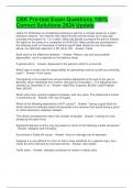
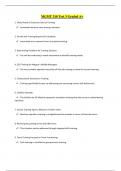
Two important models in endocrine regulation
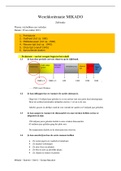
One model of endocrine regulation is the simple feedback loop,
stimulus response model or negative feedback loop (Figure 1A) and
the hierarchic control loop (Figure 1B), which is controlled by the
hypothalamus-pituitary axis.
Signal molecules can be divided into four classes
1) Peptides and proteins
-Protein structure
-Hydrophilic (do not enter cell; bind to plasma membrane)
➔ For example: ADH, oxytocin, TRH, FSH, LH, GH, PTH, α-
MSH, insulin, CCK, gastrin, ghrelin, and PRL
Figure 1. Endocrine regulation models
2) Eicosanoids (paracrine secretion) (Klaren, 2023)
-Fatty acid derivatives
-Local signalling (e.g. prostaglandins)
3) Steroids
-Cholesterol derivatives
-Produced in adrenal cortex and gonads
-Hydrophobic (enters the cell)
➔ For example: cortisol, aldosterone, estrogen and testosterone
4) Amines
-Modified amino acids
-Hydrophilic or hydrophilic, depending on size
➔ For example: epinephrine, norepinephrine (hydrophilic; both do not enter cell),
dopamine (synaptic cleft), thyroids hormones (enter cell via e.g. MCt9) (T3 & T4)
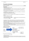
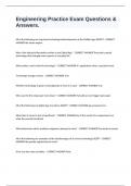
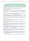
Immunohistology
Modern immunohistology useful technique in
detecting presence of hormones (Figure 2). It
starts with an antigen found in a substrate → a
primary antibody is exposed to this substrate,
attached to that this antibody is a secondary
antibody, which is bound to a (inflorescent)
reporter (HRP).
In this case HRP (a peroxidase) oxidizes DAB
(diaminobenzidine, found in benign
melanocytes), which forms DAB trimers (stain
brown under light microscopy).
Figure 2. Immunohistology overview of antibody sites (Klaren, 2023)
3
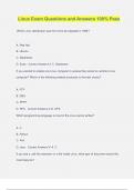
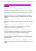
,Classical experiments in discovering hormonal pathways
A) Cori & Cori
▪ Glycogenesis (breakdown of glycogen into
glucose) stimulated by adrenaline and
glucagon
▪ Glycogen phosphorylase a (Figure 3)
breaks up glycogen into glucose polymers
▪ Adrenaline stimulated glycogenesis in
muscle but not the liver
▪ So cells can interpret signals differently by Figure 3. The role of adrenalin in activating glycogen
having different specific receptors phosphorylase beta (inactive form) (Klaren, 2023)
B) Sutherland
▪ The discovery of a second messenger (cAMP) by the use of cytoplasmatic
(glycogen phosphorylase beta; inactive) and membrane (contains adrenaline
receptors) fractions of the liver.
Complexity of cellular pathways
Cellular responses are complex, as multiple components allow for ‘fine tuning’ of response.
Signal chaining has the following different functions:
(1) Transfer of hormone signal
(2) Amplification of signal (primary signal amplification)
(3) Distribution of signal (affect processes parallel)
(4) Modulation of signal (each stap can be influenced by other factors; fine tuning!)
→ One hormone may have different functions (target cells have different receptors)
→ Intracellular response is generally a slow response (alter protein synthesis) and
extracellular cell response is faster (alter protein function)
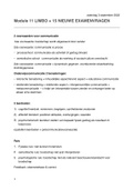
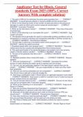
Classification of hormone receptors (Figure 4)
A) Membrane receptors
▪ Mainly for hydrophilic hormones:
peptides, catecholamines (adrenalin,
noradrenalin)
▪ Hormone binds extracellular
▪ Effect mediated via second
messengers
▪ Fast short term response (e.g.
adrenalin)
Three types of membrane (Mt) receptors:
1) Ion channel linked receptors
2) G-protein coupled receptors
3) Enzyme linked receptors (tyrosine Figure 4. Classification of hormonal receptors (Klaren, 2023)
kinases)
B) Intracellular receptors (nuclear receptors)
▪ Mainly for lipophilic (hydrophobic) hormones: steroids, thyroid hormone, calcitriol
(1,25[OH]2 – Vitamin D)
▪ Hormone receptor complex binds to DNA region
▪ Effects transcription and protein synthesis
▪ Slow long-term response (e.g. cortisol)
4
,Mt2) G-protein coupled receptors
Hormone binds to G-protein coupled
receptors (GPCR; largest group of
membrane receptors), which is coupled to
GTP binding proteins (G-proteins), which
consist of various subunits (heterotrimers):
▪ G subunits
- Gs: activates adenylate cyclase
- Gi: inhibits adenylate cyclase
- Gq: activates phospholipase C
▪ G/ subunits
The different effectors (Adenylate cyclase
and phospholipase C) produce different Figure 5. G-protein coupled receptors pathways (Klaren, 2023)
second messengers (Figure 5).
Two different G-protein transduction (effector) systems:
▪ Adenylate Cyclase
1) Converts ATP → cAMP
2) cAMP then activates Protein Kinase A (PKA)
-Protein Kinase A (PKA) phosphorylates tissue specific effectors (phosphorylase kinases,
lipases, ion channels and transcription factor CREB cycle) (!)
❌ cAMP broken down by phosphodiesterase (PDE), termination of signal
▪ Phospholipase C (PLC)
1) Converts PIP2 → diacylglycerol (DAG) + Inositol triphosphate (InsP3)
- DAG activates Protein Kinase C (PKC) → important for cell e.g. cell proliferation and
regulation of gene expression
- InsP3 binds to calcium channels and mobilises Ca2+ from intercellular stores
- Ca2+ activates: calmodulin, protein kinase C (PKC) and phospholipase A2 (PLA2)
❌ InsP3 broken down by inositol triphosphate, termination of signal
Mt3) Tyrosine kinases (Enzyme linked receptors)
Tyrosine kinases are receptor-enzyme complexes.
Tyrosine receptor inactive, monomers → Receptor active by phosphorylation, forms a dimer
Phosphorylation: addition of phosphate groups (PO4-) to OH- groups on Ser, Thr, or Tyr
residues. Regulation by:
▪ Protein kinases: catalyses phosphorylation (↑ Po4-)
▪ Phosphatases: catalyses dephosphorylation (↓ Po4-)
5
, Pathologies associated with cellular signalling
1. McCune-Albright syndrome (Figure 6)
Figure 6. Overview of McCune-Albright syndrome*
2. Cholera (Figure 7)
Affects intestine
Figure 7. Overview of cholera disease; affecting columnar epithelial cells (intestine)*
3. Androgen insensitivity syndrome (Figure 8)
Figure 8. Overview of androgen insensitivity syndrome*
4. Laron syndrome (Figure 9)
Figure 9. Overview of Laron syndrome*
6
,§2 Human histology & hormones of endocrine glands (Van der Zee)
Formation of pituitary gland (hypophysis)
Hypophyseal pouch (Rathke) + neurohypophyseal bud forms (week 3) → pouch loses
contact with roof of pharynx (month 2) → anterior + posterior formation (month 3; fetal)
Pituitary gland anatomy
The pituitary consists of two parts (Figure 10):
A) Adenohypophysis (anterior pituitary)
Consists of the following parts:
-Pars tuberalis
-Pars intermedia: MSH (Pars intermedia not present
in mammals) (Figure 11) Figure 10. Different elements of the pituitary gland (Van der Zee, 2023)
-Pars distalis
a) acidophilic cells: GH / PRL (Table 1)
b) basophilic cells: TSH / LH / FSH / ACTH
c) chromophobic cells: no granules
Adenohypophysis connected to:
-Two capillary networks (capillaries are fenestrated;
meaning that they have endothelium with pores → can
be found close to endocrine cells)
▪ Primary plexus of capillaries near hypothalamus
▪ Secondary plexus feeds the pars distalis.
-Small portals veins (Primary → Secondary possible)
B) Neurohypophysis (posterior pituitary) Figure 11. List of hormones produced and secreted in posterior and
anterior pituitary gland (Van der Zee, 2023)
Consists of the following parts:
-Pars distalis: ADH / OT (does not produce any
hormones, this is done by hypothalamus; posterior) Table 1. Cells producing the hormones in anterior pituitary
-Infundibulum
Neurohypophysis connected to:
-Capillary network
Cells present
-Non-myelinated axons from hypothalamic nerve cells
-Pituicytes (supporting glia cells, regulate secretion)
7
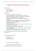
, Adrenal gland
Two adrenal glands on top of the kidney; well
vascularized
All hormones produced in adrenal glands are
close to capillaries
Adrenal gland consists of cortex and medulla
(Figure 12)
A) Cortex (Figure 13)
Zona Glomerulosa (ZG) – Stimulated by Figure 12. Different layers of the adrenal gland (Van der Zee, 2023)
angiotensin ll and potassium
- Produces Mineralo-corticoids (aldosteron) →
promote sodium reabsorption – regulates blood
pressure
Zona Fasciculata (ZF) – Stimulated by ACTH
- Spongiocytes: contain a lot of cholesterol →
Precursor of steroid hormones
- Adrenal cortex zona fasciculata cells produce
corticosteroid hormones
Figure 13. Different hormones produced by different layers of the adrenal
- Produces Gluco-Corticoids (cortisol) → Cortisol cortex (Van der Zee, 2023)
important for glucose, lipid and protein-
metabolism
Zona Reticularis (ZR) – Stimulated by ACTH
- Produces Gluco-corticoids
- Produces Androgens → Testosterone → Development of secondary characteristics and
libido
B) Adrenal medulla
Produces and secretes adrenalin and noradrenalin (fast stress hormones):
• Adrenalin: Increase of heart rate, dilation of bronchioles, dilation of arteries and
cardiac and skeletal muscle
• Noradrenaline: constriction of blood vessel of digestive system and skin, increase in
blood flow to heart, muscles and the brain
Both hormones stimulate glycogen breakdown → Increases glucose levels
8
ENDOCRINOLOGY (KW4 V)
2022-2023
Gijs Goverde (s1114555)
____________________________
,Contents
§1 Molecular mechanism of hormone signalling (Klaren) ..................................................... 3
§2 Human histology & hormones of endocrine glands (Van der Zee) .................................... 7
§3 Hypothalamus pituitary axis (Klaren) .............................................................................. 12
§4 Thyroid gland and thyroid hormones (Klaren) ................................................................. 14
§5 Growth hormone (Klaren) ............................................................................................... 18
§6 Endocrinology of adrenal glands .................................................................................... 20
§7 Endocrinology of the pancreas (Tack) ............................................................................ 26
§8 Calcitropic hormones and osteoporosis (de Vrieze)........................................................ 30
§9 Leptin and obesity (Gorissen) ........................................................................................ 34
Paraventricular (PVN) and Arcuate (ARC) nuclei of the hypothalamus ................................ 35
§10 Endocrinology of the digestive tract (Klaren) ................................................................ 37
§11 Hormones and reproduction ......................................................................................... 41
§12 Hormones and tumours (Sweep) .................................................................................. 45
Appendices ......................................................................................................................... 48
§A1 Lab protocols zebrafish ................................................................................................ 48
§A2 Results and conclusion zebrafish................................................................................. 51
§A3 Posters group C ........................................................................................................... 53
§A4 Computer class findings............................................................................................... 54
§A5 Feedback on own syllabus ........................................................................................... 55
§6 Provided feedback syllabus ............................................................................................ 57
2
,§1 Molecular mechanism of hormone signalling (Klaren)
Communication between cells
a. Endocrine (hormones)
b. Paracrine (local signaling of cell)
c. Autocrine (signaling of cell to itself)
d. Neurocrine (neurons)
e. Neuroendocrine (nerves releasing hormones)
Two important models in endocrine regulation
One model of endocrine regulation is the simple feedback loop,
stimulus response model or negative feedback loop (Figure 1A) and
the hierarchic control loop (Figure 1B), which is controlled by the
hypothalamus-pituitary axis.
Signal molecules can be divided into four classes
1) Peptides and proteins
-Protein structure
-Hydrophilic (do not enter cell; bind to plasma membrane)
➔ For example: ADH, oxytocin, TRH, FSH, LH, GH, PTH, α-
MSH, insulin, CCK, gastrin, ghrelin, and PRL
Figure 1. Endocrine regulation models
2) Eicosanoids (paracrine secretion) (Klaren, 2023)
-Fatty acid derivatives
-Local signalling (e.g. prostaglandins)
3) Steroids
-Cholesterol derivatives
-Produced in adrenal cortex and gonads
-Hydrophobic (enters the cell)
➔ For example: cortisol, aldosterone, estrogen and testosterone
4) Amines
-Modified amino acids
-Hydrophilic or hydrophilic, depending on size
➔ For example: epinephrine, norepinephrine (hydrophilic; both do not enter cell),
dopamine (synaptic cleft), thyroids hormones (enter cell via e.g. MCt9) (T3 & T4)
Immunohistology
Modern immunohistology useful technique in
detecting presence of hormones (Figure 2). It
starts with an antigen found in a substrate → a
primary antibody is exposed to this substrate,
attached to that this antibody is a secondary
antibody, which is bound to a (inflorescent)
reporter (HRP).
In this case HRP (a peroxidase) oxidizes DAB
(diaminobenzidine, found in benign
melanocytes), which forms DAB trimers (stain
brown under light microscopy).
Figure 2. Immunohistology overview of antibody sites (Klaren, 2023)
3
,Classical experiments in discovering hormonal pathways
A) Cori & Cori
▪ Glycogenesis (breakdown of glycogen into
glucose) stimulated by adrenaline and
glucagon
▪ Glycogen phosphorylase a (Figure 3)
breaks up glycogen into glucose polymers
▪ Adrenaline stimulated glycogenesis in
muscle but not the liver
▪ So cells can interpret signals differently by Figure 3. The role of adrenalin in activating glycogen
having different specific receptors phosphorylase beta (inactive form) (Klaren, 2023)
B) Sutherland
▪ The discovery of a second messenger (cAMP) by the use of cytoplasmatic
(glycogen phosphorylase beta; inactive) and membrane (contains adrenaline
receptors) fractions of the liver.
Complexity of cellular pathways
Cellular responses are complex, as multiple components allow for ‘fine tuning’ of response.
Signal chaining has the following different functions:
(1) Transfer of hormone signal
(2) Amplification of signal (primary signal amplification)
(3) Distribution of signal (affect processes parallel)
(4) Modulation of signal (each stap can be influenced by other factors; fine tuning!)
→ One hormone may have different functions (target cells have different receptors)
→ Intracellular response is generally a slow response (alter protein synthesis) and
extracellular cell response is faster (alter protein function)
Classification of hormone receptors (Figure 4)
A) Membrane receptors
▪ Mainly for hydrophilic hormones:
peptides, catecholamines (adrenalin,
noradrenalin)
▪ Hormone binds extracellular
▪ Effect mediated via second
messengers
▪ Fast short term response (e.g.
adrenalin)
Three types of membrane (Mt) receptors:
1) Ion channel linked receptors
2) G-protein coupled receptors
3) Enzyme linked receptors (tyrosine Figure 4. Classification of hormonal receptors (Klaren, 2023)
kinases)
B) Intracellular receptors (nuclear receptors)
▪ Mainly for lipophilic (hydrophobic) hormones: steroids, thyroid hormone, calcitriol
(1,25[OH]2 – Vitamin D)
▪ Hormone receptor complex binds to DNA region
▪ Effects transcription and protein synthesis
▪ Slow long-term response (e.g. cortisol)
4
,Mt2) G-protein coupled receptors
Hormone binds to G-protein coupled
receptors (GPCR; largest group of
membrane receptors), which is coupled to
GTP binding proteins (G-proteins), which
consist of various subunits (heterotrimers):
▪ G subunits
- Gs: activates adenylate cyclase
- Gi: inhibits adenylate cyclase
- Gq: activates phospholipase C
▪ G/ subunits
The different effectors (Adenylate cyclase
and phospholipase C) produce different Figure 5. G-protein coupled receptors pathways (Klaren, 2023)
second messengers (Figure 5).
Two different G-protein transduction (effector) systems:
▪ Adenylate Cyclase
1) Converts ATP → cAMP
2) cAMP then activates Protein Kinase A (PKA)
-Protein Kinase A (PKA) phosphorylates tissue specific effectors (phosphorylase kinases,
lipases, ion channels and transcription factor CREB cycle) (!)
❌ cAMP broken down by phosphodiesterase (PDE), termination of signal
▪ Phospholipase C (PLC)
1) Converts PIP2 → diacylglycerol (DAG) + Inositol triphosphate (InsP3)
- DAG activates Protein Kinase C (PKC) → important for cell e.g. cell proliferation and
regulation of gene expression
- InsP3 binds to calcium channels and mobilises Ca2+ from intercellular stores
- Ca2+ activates: calmodulin, protein kinase C (PKC) and phospholipase A2 (PLA2)
❌ InsP3 broken down by inositol triphosphate, termination of signal
Mt3) Tyrosine kinases (Enzyme linked receptors)
Tyrosine kinases are receptor-enzyme complexes.
Tyrosine receptor inactive, monomers → Receptor active by phosphorylation, forms a dimer
Phosphorylation: addition of phosphate groups (PO4-) to OH- groups on Ser, Thr, or Tyr
residues. Regulation by:
▪ Protein kinases: catalyses phosphorylation (↑ Po4-)
▪ Phosphatases: catalyses dephosphorylation (↓ Po4-)
5
, Pathologies associated with cellular signalling
1. McCune-Albright syndrome (Figure 6)
Figure 6. Overview of McCune-Albright syndrome*
2. Cholera (Figure 7)
Affects intestine
Figure 7. Overview of cholera disease; affecting columnar epithelial cells (intestine)*
3. Androgen insensitivity syndrome (Figure 8)
Figure 8. Overview of androgen insensitivity syndrome*
4. Laron syndrome (Figure 9)
Figure 9. Overview of Laron syndrome*
6
,§2 Human histology & hormones of endocrine glands (Van der Zee)
Formation of pituitary gland (hypophysis)
Hypophyseal pouch (Rathke) + neurohypophyseal bud forms (week 3) → pouch loses
contact with roof of pharynx (month 2) → anterior + posterior formation (month 3; fetal)
Pituitary gland anatomy
The pituitary consists of two parts (Figure 10):
A) Adenohypophysis (anterior pituitary)
Consists of the following parts:
-Pars tuberalis
-Pars intermedia: MSH (Pars intermedia not present
in mammals) (Figure 11) Figure 10. Different elements of the pituitary gland (Van der Zee, 2023)
-Pars distalis
a) acidophilic cells: GH / PRL (Table 1)
b) basophilic cells: TSH / LH / FSH / ACTH
c) chromophobic cells: no granules
Adenohypophysis connected to:
-Two capillary networks (capillaries are fenestrated;
meaning that they have endothelium with pores → can
be found close to endocrine cells)
▪ Primary plexus of capillaries near hypothalamus
▪ Secondary plexus feeds the pars distalis.
-Small portals veins (Primary → Secondary possible)
B) Neurohypophysis (posterior pituitary) Figure 11. List of hormones produced and secreted in posterior and
anterior pituitary gland (Van der Zee, 2023)
Consists of the following parts:
-Pars distalis: ADH / OT (does not produce any
hormones, this is done by hypothalamus; posterior) Table 1. Cells producing the hormones in anterior pituitary
-Infundibulum
Neurohypophysis connected to:
-Capillary network
Cells present
-Non-myelinated axons from hypothalamic nerve cells
-Pituicytes (supporting glia cells, regulate secretion)
7
, Adrenal gland
Two adrenal glands on top of the kidney; well
vascularized
All hormones produced in adrenal glands are
close to capillaries
Adrenal gland consists of cortex and medulla
(Figure 12)
A) Cortex (Figure 13)
Zona Glomerulosa (ZG) – Stimulated by Figure 12. Different layers of the adrenal gland (Van der Zee, 2023)
angiotensin ll and potassium
- Produces Mineralo-corticoids (aldosteron) →
promote sodium reabsorption – regulates blood
pressure
Zona Fasciculata (ZF) – Stimulated by ACTH
- Spongiocytes: contain a lot of cholesterol →
Precursor of steroid hormones
- Adrenal cortex zona fasciculata cells produce
corticosteroid hormones
Figure 13. Different hormones produced by different layers of the adrenal
- Produces Gluco-Corticoids (cortisol) → Cortisol cortex (Van der Zee, 2023)
important for glucose, lipid and protein-
metabolism
Zona Reticularis (ZR) – Stimulated by ACTH
- Produces Gluco-corticoids
- Produces Androgens → Testosterone → Development of secondary characteristics and
libido
B) Adrenal medulla
Produces and secretes adrenalin and noradrenalin (fast stress hormones):
• Adrenalin: Increase of heart rate, dilation of bronchioles, dilation of arteries and
cardiac and skeletal muscle
• Noradrenaline: constriction of blood vessel of digestive system and skin, increase in
blood flow to heart, muscles and the brain
Both hormones stimulate glycogen breakdown → Increases glucose levels
8