100% satisfaction guarantee Immediately available after payment Both online and in PDF No strings attached
logo-home
Understand the importance of microbial classification to medicine and industry. Undertake microscopy for specimen examination in laboratories. CA$31.94   Add to cart

Essay

Understand the importance of microbial classification to medicine and industry. Undertake microscopy for specimen examination in laboratories.

 11 views  0 purchase
  • Course
  • Institution

A research report using any appropriate format that covers four of the listed microorganisms. Practical work setting up and using light microscopes and oil immersion lenses to look at the structure of microorganisms. Scientific drawings of specimens, laboratory notebooks and practical write-ups sup...

[Show more]
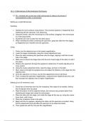
Preview 3 out of 24  pages

  • August 12, 2024
  • 24
  • 2023/2024
  • Essay
  • Unknown
  • A+
avatar-seller
16 A + B Microbiology & Microbiological Techniques

P2 - Correctly set up and use a light microscope to observe structures of
microorganisms under a microscope:

Setting up a Light Microscope:

Handling:

1. Sanitise the work surfaces using alcohol. This prevents bacteria or fingerprints from
interfering with the specimen I am observing.
2. Using two hands, carry the microscope to a flat surface, plugging in the microscope
to a near plug socket
3. Guarantee the cord is away from the desk space
4. When handling the slides containing the specimen, grasp the slide from the edges,
so fingerprints don’t transfer onto the specimen.

Using:

1. Firstly, turn the objective lens to the lowest magnification.
2. Lower the stage considerably, using the coarse adjustment knob.
3. Place the slide containing the specimen onto the stage, aligning it with the circular
hole in the stage.
4. Make sure to secure the stage clips onto the end of each edge of the slide, to hold it
into place.
5. Observe the specimen through the eyepiece to determine if it needs adjusting to be
more central.
6. Using the coarse adjustment knob, raise the stage, and turn on the light.
7. Next, observe through the eyepiece lens and adjust the distance o the stage from the
objective lens
8. Once the specimen is in focus, use the fine adjustment knob to add focus
9. Then, increase the specimen magnification, using the lower objective lens (4x), and
then increase the objective lens to higher magnification

Setting up an Oil Immersion Lens:

1. Screw the oil immersion lens into the nosepiece, then adjust it as needed, clicking
the oil objective lens into place
2. Wipe the bottom of all the objective lenses, this removes any present debris from
disturbing the image seen under the microscope
3. Place the slide onto the stage, and rotate the stage all the way up, using the coarse
focus knob, until it stops
4. Place the condenser close to the slide
5. Begin with the 4x eyepiece, adjusting the state until the specimen is located. Using
the fine adjustment knob will allow the specimen to be observed clearly
6. Then repeat this with the 10x and 40x magnification.

, 7. Slide the 40x slightly out of the way to allow a drop of immersion oil onto the cover
slip of the specimen
8. The using the 100x oil immersion lens and use the fine focus knob to observe the
specimen clearly

M2 - Compare the use of different Microscopy techniques to observe the structures of
microorganisms

Microscopy:

Light microscope

In a light microscope, visible light passes through the specimen, this is then bent through the
lens system. Therefore, allowing the person to see a magnified image of the specimen. Light
microscopy can be used on living cells, so analysing cell behaviours is possible under this
microscope. This type of microscopy uses a single convex lens of a small focal length for
magnification. Usually the magnification can reach up to x1500, though this is uncommon in
light microscopes used in school educational settings. With a light microscope, studies on
specimens like algae, fungi and insects can be made. In addition, light microscopes can be
used to study soil types, cells, bacteria, blood samples and hair follicles. Some organelles of
a cell can be seen such as the cell membrane, mitochondria and chloroplasts.

Compound light microscope

A compound microscope functions when light is focused through its condenser on the stage.
The light transmitted by the specimen is then shown through the objective lens. This creates
a magnified image called the primary image. Then the light bends magnifying the image,
creating a highly double magnified image. The magnification of a compound light microscope
ranges between 40x to 1000x. In order to determine the magnification, use the power of the
objective lens and multiply it by the eyepiece power, this will give you the total magnification.
Compound microscopes can be used in many circumstances such as the morphology of
microorganisms and cellular structure. Under a compound light microscope, red blood cells
can be seen under great detail, to show the platelets. Additionally, bacteria can be easily
viewed and studied under compound light microscopes. Other images can be seen such as
microorganisms, tissue samples, and cellular components such as the mitochondria etc.

Phase contrast microscope

Phase contrast microscope is an optical microscope. The light from the illuminator focuses
on the specimen being looked at. Then, the light passes through various regions of the slide,
creating different refractive indexes and thicknesses. An optical device is used to convert
phase shifts. The brightness change is then seen, creating observable contrast in the final
image. There are many uses to phase contrast microscopes, one being they observe living
cells in their natural form. Additionally, it allows an observation of protozoans, diatoms and
planktons. Therefore, it can be used to study subcellular structures and cellular processes. A
compound microscope allows magnification between 4x to 100x.

, Inverted microscope

An inverted microscope is a form of light microscope. Using the objective lenses and turret
below its stage and the illuminator and condenser above the stage, allows the images to be
seen through the eyepiece lenses. With this microscope, you have to look upwards to view
the specimen being studied. There are many uses for an inverted microscope. For example,
you can observe metallurgical processes of metals and minerals, cytology of cell division
and detection of tuberculosis in culture. The magnification observed under an inverted
microscope is between 40x to 400x.

Electron microscope SEM (scanning electron microscope) & TEM (transmission
electron microscope)

An electron microscope uses accelerated electrons rather than light rays to illuminate a
specimen, and produce a highly magnified image. Due to the extremely short electron
wavelengths, the microscope allows very high resolution of the image, along with
magnification up to 10,000,000x. As a result, high quality images with high contrast can
reveal detailed structures. A specimen up to 0.2nm can be seen under electron
microscopes. All types of electron microscopes allow observations of virus, flagella, pili and
bacterial cell structure. Additionally, it can be used in forensic to study ballistics and drug
development. Lastly, it can be used in geology to study minerals and rocks.

Comparison of how using two types of Microscopes is important and helpful when
viewing the structure of microorganisms:

In order for an observer to view the structure of a microorganism, it may be best to use a
compound light microscope or an electron microscope. Which microscope they use will
depend on what they aim to view about the specimen. This is due to the different operating
principles of the microscopes. They produce completely different images for the observer
depending on what they wish to view specifically.

Compound light microscopes use visible light and magnifying lenses to observe specimens.
The light travels through the eyepiece and objective lenses with the maximum magnification
of 1500x and resolution of 200 nm. However, because the wavelength of visible light is too
large to pass between small cell organelles, they cannot be seen or analysed. Light
microscopes allow the observations of algae, fungi, cells, bacteria, blood and hair follicles.
Additionally, the larger organelles, such as cell membrane, mitochondria and chloroplasts
can be seen, however, finer organelles are impossible to be identified. The lack of
observation for smaller organelles is a major disadvantage of light microscopes. Many
microorganisms may not be viewed, due to the light microscope offering low magnification
and resolution, which I previously mentioned. However, as light microscopes operate using
visible light, it allows the observer to view living and moving specimens, rather than killing
the specimen due to any high radiation levels (what occurs when electron microscopes are
used). Light microscopes are incredibly cost effective, its basic design allows it to be
relatively low cost. Additionally, they do not require extensive training to operate the
equipment so many academic facilities use light microscopes.

The benefits of buying summaries with Stuvia:

Guaranteed quality through customer reviews

Guaranteed quality through customer reviews

Stuvia customers have reviewed more than 700,000 summaries. This how you know that you are buying the best documents.

Quick and easy check-out

Quick and easy check-out

You can quickly pay through credit card or Stuvia-credit for the summaries. There is no membership needed.

Focus on what matters

Focus on what matters

Your fellow students write the study notes themselves, which is why the documents are always reliable and up-to-date. This ensures you quickly get to the core!

Frequently asked questions

What do I get when I buy this document?

You get a PDF, available immediately after your purchase. The purchased document is accessible anytime, anywhere and indefinitely through your profile.

Satisfaction guarantee: how does it work?

Our satisfaction guarantee ensures that you always find a study document that suits you well. You fill out a form, and our customer service team takes care of the rest.

Who am I buying these notes from?

Stuvia is a marketplace, so you are not buying this document from us, but from seller severino6. Stuvia facilitates payment to the seller.

Will I be stuck with a subscription?

No, you only buy these notes for CA$31.94. You're not tied to anything after your purchase.

Can Stuvia be trusted?

4.6 stars on Google & Trustpilot (+1000 reviews)

75323 documents were sold in the last 30 days

Founded in 2010, the go-to place to buy study notes for 14 years now

Start selling
CA$31.94
  • (0)
  Add to cart