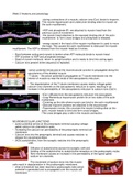
100% satisfaction guarantee Immediately available after payment Both online and in PDF No strings attached
logo-home
notes on joints and muscles £9.29
Add to cart

Lecture notes

notes on joints and muscles

 0 purchase

notes on muscles and joints fully summarised with pictures

Preview 1 out of 4  pages

  • May 16, 2021
  • 4
  • 2020/2021
  • Lecture notes
  • Dr sheila
  • Muscles
book image

Book Title:

Author(s):

  • Edition:
  • ISBN:
  • Edition:
All documents for this subject (1)
avatar-seller
fathimafareiz
Week 5 Anatomy and physiology

-during contractions of a muscle, calcium ions (Ca+) binds to troponin.
This moves tropomyosin and undercover binding sites for myosin on
the actin myo lament.

-ADP and phosphate (P) are attached to myosin head from the
previous cycle of movement.
-the myosin head attaches to the exposed binding site of the actin
myo lament to form cross bridges and phosphate is released.

-energy stored in the head of the myosin myo lament is used to move
the head. This causes the actin myo lament to slide past the myosin
myo lament. The ADP is released from the myosin head as it moves.

- Bond between acting and myosin is broken when ATP molecule binds to myosin head
- ATP is broken to ADP and phosphate (energy)
- Head of myosin molecule return to upright position and is ready to bind into acting again.
- Calcium Ions present entire sequence is repeated.


-action potential introduced at the neuromuscular junction is propagated along the
sarcolemma of the skeletal muscle.
-T tubule - the action potential is propagated on T tubule membrane into the
interior of muscle so near the ends of the sarcoplasmic reticulum.
-
- The depolarisation of the T tubule membrane causes nearby voltage gated
calcium ions channels on the sarcoplasmic reticulum to open, resulting in an
increase in the permeability of the sarcoplasmic reticulum to calcium ions (Ca2+).

-(Ca+) di uses from the sarcoplasmic reticulum into sarcoplasm.
-Long lamentous tropomyosin protein lie on two sides of the actin
mo lament.
-Covering up the site where myosin can bind to the actin myo lament
-Globular troponin proteins are attached to the tropomyosin
-Tropomyosin moves, this uncovers the myosin binding sites on the
actin, myosin heads to bind and form cross bridges.
-The cross bridges play a role in the muscle contraction

NEUROMUSCULAR JUNCTIONS
- action potential arrives at the presynaptic terminal causing voltage
gated calcium ion channels to open,
- Increasing the calcium ion permeability of the presynaptic terminal cell
membrane
- Calcium ions into the presynaptic terminal and causes vesicles to
release the neurotransmitters
- (ACh) acetylcholine, from the synaptic vesicles into the presynaptic
cleft.

Di usion of acetylcholine across the synaptic cleft and
bidding of the acetylcholine to acetylcholine receptors on the postsynaptic mules
bre membrane causes an increase in the permeability of the ligand-gated
sodium ion channels.

The movement of sodium irons into the muscle
cells result in depolarisation in the postsynaptic membrane.
- what is threshold has been reached a postsynaptic take action
potential is generated and is propagated over the muscle cell
membrane


fi fffifi fi ff

fi fi fi fi

The benefits of buying summaries with Stuvia:

Guaranteed quality through customer reviews

Guaranteed quality through customer reviews

Stuvia customers have reviewed more than 700,000 summaries. This how you know that you are buying the best documents.

Quick and easy check-out

Quick and easy check-out

You can quickly pay through credit card for the summaries. There is no membership needed.

Focus on what matters

Focus on what matters

Your fellow students write the study notes themselves, which is why the documents are always reliable and up-to-date. This ensures you quickly get to the core!

Frequently asked questions

What do I get when I buy this document?

You get a PDF, available immediately after your purchase. The purchased document is accessible anytime, anywhere and indefinitely through your profile.

Satisfaction guarantee: how does it work?

Our satisfaction guarantee ensures that you always find a study document that suits you well. You fill out a form, and our customer service team takes care of the rest.

Who am I buying these notes from?

Stuvia is a marketplace, so you are not buying this document from us, but from seller fathimafareiz. Stuvia facilitates payment to the seller.

Will I be stuck with a subscription?

No, you only buy these notes for £9.29. You're not tied to anything after your purchase.

Can Stuvia be trusted?

4.6 stars on Google & Trustpilot (+1000 reviews)

68175 documents were sold in the last 30 days

Founded in 2010, the go-to place to buy revision notes and other study material for 15 years now

Start selling
£9.29
  • (0)
Add to cart
Added