Electrical Activity in the Heart
7.8 i) Know the myogenic nature of cardiac muscle.
ii) Understand how the normal electrical activity of the heart coordinates the heart beat, including the
roles of the sinoatrial node (SAN), the atrioventricular node (AVN), the bundle of His and the
Purkyne fibres.
Cardiac Muscle
• Cardiac muscle is myogenic. Electrical activity in the heart creates rhythmic patterns of contractions,
which coordinates the regular heart beat.
• Myogenic: Muscle that can contract without external nervous stimulation.
Control of the Heart Beat
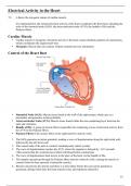
• Sinoatrial Node (SAN): Muscle tissue found in the wall of the right atrium, which acts as a
pacemaker and generates action potentials.
• Atrioventricular Node (AVN): Muscle tissue found within the non-conducting layer between the
atria and ventricles.
• Bundle of His: A group of muscle fibres responsible for conducting waves of electrical activity from
the AVN to the Purkyne fibres.
• Purkyne Fibres: Fine muscle fibres in the right and left ventricle walls.
1. The SAN generates an action potential, sending a wave of depolarisation along the right atrial wall,
followed by the left atrial wall.
2. This causes both of the atria to contract simultaneously (atrial systole).
3. The wave of depolarisation reaches the AVN, where the impulse is delayed by ~0.13 seconds.
(This ensures that the ventricles have filled with blood before contraction)
4. The wave of depolarisation then travels to the apex of the heart via the bundle of His.
5. The impulse spreads up through the Purkyne fibres into the ventricle walls, causing the muscle to
2
contract from the base upwards (ventricular systole).
6. Blood is forced into the arteries and there is a short delay before the next action potential is
generated, during which time the heart muscles relax and repolarise (diastole).
13