100% satisfaction guarantee Immediately available after payment Both online and in PDF No strings attached
logo-home
SUMMARISED OCR A-Level Biology Module 5 Notes, based on Mark Schemes £9.66
Add to cart

Other

SUMMARISED OCR A-Level Biology Module 5 Notes, based on Mark Schemes

 14 views  0 purchase
  • Institution
  • OCR

These notes got me an A* in OCR A-Level Biology. These Module 5 notes were made to be bite-sized and straight to the point. Best part? They incorporate MARK SCHEME answers from previous OCR past paper questions so you can get an exact idea of what the exam wants from you. There's also a column for ...

[Show more]
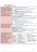
Preview 4 out of 31  pages

  • June 19, 2024
  • 31
  • 2023/2024
  • Other
  • Unknown
All documents for this subject (12)
avatar-seller
SM12345
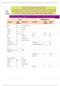
SPEC MODULE 5
5.1.1 Communication and Homeostasis
(a) the need for communication Why multicellular need communication systems:
systems in multicellular organisms ● Small SA:V (1) diffusion distance is too large (1) e.g to transport sucrose
To include the need for animals and from source to sink (1)
plants to respond to changes in the Animals and plants need to respond to changes in internal & external
internal and external environment environments:
and to coordinate the activities of ● Internal: e.g blood glucose conc, internal body temp,
different organs. ● External: temp, light intensity, humidity, water potential, pH

(b) the communication between cells State what is meant by cell signalling. (1)
by cell signalling. To include ● Communication between cells
signalling between adjacent cells ● If 2 marks, add → to trigger reaction/ response
and signalling between distant cells. Signalling can occur between adjacent cells (e.g callose signals to neighbouring
cells of pathogen attack) OR
Between distant cells (e.g signalling molecule travels in blood to the target cell)

Explain how cell surface membranes contribute to the process of cell
signalling. (4)
● Signal molecule released by exocytosis
● Glycoproteins have receptors, which are specific
● Shape of receptor and the signal molecule is complementary
● 4th mark comes from key vocab

(c) the principles of homeostasis To What is meant by homeostasis? (3)
include the differences between ● Maintaining a stable internal environment (1) within a narrow range (1)
receptors and effectors, and the even though the environment is changing (1)
differences between negative
Receptors: detect the stimuli
feedback and positive feedback.
Effectors: coordinate the response
Negative feedback: counteracts initial change and restores it to the normal level
(e.g blood glucose conc) ← is homeostasis
Positive feedback: initial change is exaggerated (1) (e.g blood clotting,
hypothermia) ← NOT homeostasis

(d) the physiological and
behavioural responses involved in
Physiological Behavioural
temperature control in ectotherms
and endotherms. Ectotherms - - Dark colours → absorb - Increase or decrease
To include, endotherms – peripheral depend on more radiation radiation absorbed (e.g lying in
temperature receptors, the role of external - Alters heart rate → can the sun)
the hypothalamus and effectors in environment reduce metabolic rate - Minimise movement
skin and muscles; behavioural
responses, ectotherms – Endotherms - - Peripheral body temp - Hibernation → reduces
behavioural responses. body temp receptors metabolic rate → keeps warm
An opportunity to monitor controlled by - Thermoregulatory centres - E.g flap ears to cool down
physiological functions in homeostasis - High metabolic rate to
ectotherms and/or endotherms. maintain internal body temp

,Describe the way in which an Mammals:
endothermic animal, such as a ● Thermoreceptors stimulated by changes in blood temperature (1)
mammal, normally prevents its ● Impulses sent to the hypothalamus (1)
body temperature from ● Then…
decreasing when the external
Cooling Down Warming Up
temperature decreases. (5)
● Thermoreceptors stimulated ● Vasodilation ● Vasoconstriction of arterioles
by changes in blood Vessels near skin dilate so more Vessels near skin constrict so less
temperature (1) blood flow so more heat lost by blood flow so less heat lost by
● Impulses sent to the radiation radiation
hypothalamus (1) ● Hairs lie flat ● Hairs raise and trap air
● Vasoconstriction of arterioles So they don’t trap insulating air Traps layer of insulating air
(1) reducing heat loss by ● Sweating ● Shivering to generate heat
radiation (1) Heat lost through water evaporating Contracting & relaxing of skeletal
● Shivering to generate heat as water has a high specific latent of muscles generate metabolic heat
(1) vaporisation
● Hairs raise and trap air (1)
Thermoreceptors send impulses along sensory neurones to the hypothalamus,
which sends impulses along the motor neurones to the effector (e.g skeletal
muscles/ erector pili muscles)
5.1.2 Excretion as an example of homeostatic control
(a) the term excretion and its Excretion: removal of waste products of metabolism from the body (2)
importance in maintaining Metabolism: all the chemical reactions that happen in a cell/ organism
metabolism and homeostasis To Importance of excretion:
include reference to the importance ● Avoid the buildup of products which could be toxic
of removing metabolic wastes, E.g too much CO2 → pH decreases → Acidosis
including carbon dioxide and E.g too much ammonia → pH increases → Alkalosis
nitrogenous waste, from the body. E.g too much urea → water potential decreases
E.g too much bile → jaundice

(b) (i) the structure and functions of i) Function: detox (breaks down metabolic products into less harmful products),
the mammalian liver glycogen store, forms urea
(ii) the examination and drawing of External Structure:
stained sections to show the Hepatic artery: brings oxygenated blood
histology of liver tissue Hepatic vein: removes deoxygenated blood
To include the gross structure and Hepatic portal vein: brings deoxygenated
histology of the liver AND the roles blood from the duodenum, carrying
of the liver in the storage of digestion products
glycogen, detoxification and the Gall bladder: stores bile
formation of urea (the ornithine cycle
covered in outline only). Internal Structure:
made up of many lobules, which contain:
Roles of liver: Hepatocytes: liver cells (have large nuclei
● Detoxification and a lot of mitochondria)
● Formation of urea Sinusoids: where H artery and H portal vein mix, increasing the oxygen content
● Glycogen storage Hepatocytes break down harmful → less harmful stuff, which re-enters blood
(glycogenesis: glucose to Kupffer Cells: macrophages remove bacteria & prevent disease & break down
glycogen) old RBCs
Bile Canaliculi: where bile produced by hepatocytes is secreted to

, Bile duct: where bile drains off into the gall bladder


Formation of Urea: liver also breaks down excess amino acids
1) Deamination: removing amine group to form ammonia (and organic acid)
However, ammonia is toxic so…
Ornithine Cycle: 2) Ornithine cycle converts ammonia to urea ( NH3 + CO2 → urea + H2O)

Ornithine + CO2 + NH3 → citruline + H2O (in mitochondria)
Citruline + NH3 → Arginine + H2O
Arginine + H2O → Ornithine + urea

Detoxification: breakdown of harmful substances into less harmful stuff. E.g…
1) Alcohol: ethanol → ethanal → acetic acid (less harmful)
Excess alcohol over time leads to cirrhosis, where liver cells die
2) Paracetamol: painkiller broken down in the liver
Excess paracetamol can lead to liver and kidney failure
3) Insulin:
May involve active transport as Excess insulin can lead to problems with blood glucose levels
diffusion [of substances around the
cycle] is too slow (2) Glycogen storage: liver convert excess glucose into glycogen (glycogenesis) →
stored as granules in liver cells → released when needed

(c) (i) the structure, mechanisms of External Structure:
action and functions of the ● Blood flows through renal artery and
mammalian kidney removed by renal vein
(ii) the dissection, examination and ● Cortex - dark outer layer → filters blood +
drawing of the external and internal
has capillaries from renal artery to the
structure of the kidney
(iii) the examination and drawing of nephrons
stained sections to show the ● Medulla - light → contains tubules of the
histology of nephrons nephrons (form pyramids) + collecting
To include the gross structure and ducts
histology of the kidney including the ● Pelvis: lighter
detailed structure of a nephron and In the nephron:
its associated blood vessels AND
1) Ultrafiltration
the processes of ultrafiltration,
selective reabsorption and the ● Glomerulus - network of capillaries
production of urine. ● Afferent arterioles takes blood into glomerulus and efferent
arterioles take blood out of the glomerulus

, ● Afferent arteriole has a greater diameter than efferent arteriole (1)
Ultrafiltration: afferent & efferent high hydrostatic pressure (1) [High pressure forces small
arteriole, high hydrostatic pressure, molecules into the Bowman’s Capsule but large molecules remain
endothelium walls, basal membrane,
in the blood]
podocytes
● Endothelium wall has small pores for passage (1)
● Basal membrane stops large molecules (1)
● Podocytes ensures there’s gaps (1)
● They pass through the capillary endothelium to the basement
membrane to the epithelium of the Bowman’s capsule
2) Selective Reabsorption
● Filtrate flows through the PCT, Loop of Henle, DCT and collecting duct
and stuff (e.g glucose, AAs, H2O) are reabsorbed into the blood
PCT:
Has microvilli (large SA) for reabsorption of glucose, AAs, vitamins etc
● Na+ is pumped into the PCT by active transport, causing a higher conc in
the PCT
● Na+ moves down the conc gradient, while cotransporting a molecule (e.g
glucose) from filtrate to the ciliated cells
● Glucose & AAs enter blood by facilitated diffusion
Cortex: Loop of Henle: decreases the water potential
● Filtrate leaving PCT is very concentrated
● In the ascending limb, ions are actively transported out [ → medulla
tissue fluid is very concentrated] & is NOT permeable to water
● In the descending limb, water moves out by osmosis [down conc gradient
into the concentrated medulla tissue fluid] & is permeable to water
DCT:
Medulla:
● Further reabsorption of water & ions
● Cotransport in DCT only involves ions
Collecting duct:
● Passes through conc medulla tissue fluid
● Depending on ADH levels, water can diffuse out, down conc gradient
Urine:
● Filtrate remaining passes through the ureter to the bladder
● Contains no proteins (too large), glucose (all reabsorbed)
● Contains urea, excess vitamins, hormones, water etc
External Structure:


ii) External and internal structures from dissection

The benefits of buying summaries with Stuvia:

Guaranteed quality through customer reviews

Guaranteed quality through customer reviews

Stuvia customers have reviewed more than 700,000 summaries. This how you know that you are buying the best documents.

Quick and easy check-out

Quick and easy check-out

You can quickly pay through credit card for the summaries. There is no membership needed.

Focus on what matters

Focus on what matters

Your fellow students write the study notes themselves, which is why the documents are always reliable and up-to-date. This ensures you quickly get to the core!

Frequently asked questions

What do I get when I buy this document?

You get a PDF, available immediately after your purchase. The purchased document is accessible anytime, anywhere and indefinitely through your profile.

Satisfaction guarantee: how does it work?

Our satisfaction guarantee ensures that you always find a study document that suits you well. You fill out a form, and our customer service team takes care of the rest.

Who am I buying these notes from?

Stuvia is a marketplace, so you are not buying this document from us, but from seller SM12345. Stuvia facilitates payment to the seller.

Will I be stuck with a subscription?

No, you only buy these notes for £9.66. You're not tied to anything after your purchase.

Can Stuvia be trusted?

4.6 stars on Google & Trustpilot (+1000 reviews)

52510 documents were sold in the last 30 days

Founded in 2010, the go-to place to buy revision notes and other study material for 14 years now

Start selling
£9.66
  • (0)
Add to cart
Added