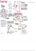
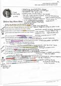
The heart- blood Artery Vein
captain
[ Head-neck [
I
lumen
O
G veinscontainvaluestoea
s
Hi
lumens
L
Endothelium >
4
<
Lining
e10
Upper limbs < -
↓ friction
) # (h)
I
Pulmonary
d
artery Pulmonary Elastic layer Elastic in veins there is lower
vein
maintain layer , a
>
Lungs J -
· layed
TransportSystems Vena V
pressure so muscle contraction
Cava in surrounding tissues his
·
transport medium that moves
> R Atrium L Atrium <
&
. .
in bulk in a continuous system -
↓
~
to move the blood
of tubular vessels
.
R Ventricle Aorta
L Ventricle
.
M 2
Veins also have thin walls
M
allowing external
Hepatic
E [
vein
Liver
Hepatic artery
Tough outer muscles to
'squeze' and they are able to distend
I
[
collagen fibres
·
Aorta easily to accomodate changing volumes of blood
O
A
s
j Intestines S
S &
jj
Pulmonary artery
&
Renal vein Renal sallows formation of tissue fluid
artery
&S
- >
Kidneys
pulmonary value Pulmonary vein
IS
↓ E
↓
L
eaveEfyoo
&Na > Lower l i m b s
rightatrin
Left atrium hydrostatic
00 80
hydrostatic
S
·
↑ pressure ↓ pressure
I &
X
l
Bicuspid mitral value
Humans have
system where blood
a double circulatory
flows through plasmad
A &
11 CO2
↓d
-proteins
N&
that remain
Chordae tendinae ultrafiltration
·
A single circulatory ions
h
the heart twice
Rig ht ventricle Left ventricle #
&
.
Urea
glucose
&Y
system has I ventricle and blood is
salts
amino a
I Lymph drainsintobloodaas
·
·un Sa
>
4
-
Interventricular .
mixed
pericardinium
j&
N
A
septum a
/
W- N Thick because blood L
4
fluid)
mines a
·
reda
from Lu goes further
exess
than that from RV
Apex tissue
↓ contains
fatty acids
·
SA node releases an electrical impulse
+ fats S
&
relaxing ↳ sel L
·impulsetraves downatriatheycontract Crelaxingasing relaxing Cardiovascular disease
↓
&
Catherosclerosis)
&) damaged
0 endothelial cells
!
down interventricular septum eup
2
around
*
L
& -
macrophage WBC respond
close L
inflamation
↓
&Ch >
-
T
ventricles them to contract.
causing blood - rC ↳ forms
&
aorta g
walls stretch
moving - Low density lipoproteins foam cells
away from
heart ↳ designed to bring
High density lipoproteins
>
&
>
Aortic pressure ↳ designed to cholesterd to the cells
carrycholester
at
to the liver
ventricular pressure &
0%
00
%
- y
restricted
~
Av
opens
Op C
- s
· ↑
11 blood flow
Atrial pressure
8
11 -1
&-
(
filling
·
6
e atria
ventricles
080
& 0
--
↑ pressure => anyeurism
Platlets = clotting = thrombosis