Whether you're a radiography student, a healthcare professional, or simply interested in medical imaging, this content will provide you with a comprehensive understanding of soft tissue radiography and its role in modern healthcare
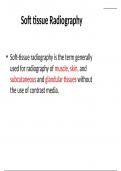
• Soft-tissue radiography is the term generally
used for radiography of muscle, skin, and
subcutaneous and glandular tissues without
the use of contrast media.
, Exposure technique
• Several different exposure techniques may be
used.
• They may be divided broadly into two
categories: those employing a
1. Normal kVp
2. Non-standard kVp.
, Normal kVp
This category may be divided into three
subcategories:
• Use of a normal technique for the part being
examined when air shadows or fat pads may
delineate abnormalities in adjacent soft tissue
structures, e.g.: – effusion in a synovial cavity
causing a filling defect in a fat pad adjacent to
a joint; – enlarged adenoids causing a filling
defect in the air contained in the nasopharynx.
,• Use of two or more films or film/screen
combinations to demonstrate both bony detail and
soft tissue with one exposure, e.g.: – facial bones,
nasal bones and soft tissues of the face; –
calcification of tendons and bony detail of the
shoulder joint.
• Use of a wedge filter, where the thicker part of the
wedge attenuates the beam over the soft tissues,
e.g.: – cephalography to demonstrate bony detail of
the skull and facial bones along with the soft-tissue
outline of the face on one film; – to see the soft
tissue of the toes when exposing the whole foot.
, Non standard kVp
• This category of exposure technique may also
be divided into three subcategories:
1. subnormal kVp,
2. low kVp and
3. high kVp.
, Subnormal kVp
• This term is used when the kVp employed in less than 45
kVp, which is the lowest useful kVp available on many X-
ray units.
• Modified or special equipment is required that has an X-
ray tube with reduced added and inherent filtration
along with a small focal spot size.
• The use of such kVp increases differential attenuation of
adjacent soft tissues and thus increases subject contrast.
• An example of the use of this technique is found in
mammography.
The benefits of buying summaries with Stuvia:
Guaranteed quality through customer reviews
Stuvia customers have reviewed more than 700,000 summaries. This how you know that you are buying the best documents.
Quick and easy check-out
You can quickly pay through credit card for the summaries. There is no membership needed.
Focus on what matters
Your fellow students write the study notes themselves, which is why the documents are always reliable and up-to-date. This ensures you quickly get to the core!
Frequently asked questions
What do I get when I buy this document?
You get a PDF, available immediately after your purchase. The purchased document is accessible anytime, anywhere and indefinitely through your profile.
Satisfaction guarantee: how does it work?
Our satisfaction guarantee ensures that you always find a study document that suits you well. You fill out a form, and our customer service team takes care of the rest.
Who am I buying these notes from?
Stuvia is a marketplace, so you are not buying this document from us, but from seller sathyapriyathanarasuu. Stuvia facilitates payment to the seller.
Will I be stuck with a subscription?
No, you only buy these notes for £3.58. You're not tied to anything after your purchase.