Part 1: Introduction and Background Information
https://www.ncbi.nlm.nih.gov/pmc/articles/PMC4486472/pdf/emss-63689.pdf = review
Cause of NTD: result from failure of fusion of the neural tube during early embryogenesis
Common CNS malformations
- Affect 1 per 1000 pregnancies
- Over 250, 000 cases per year
Main Types of NTDs
1. Anencephaly (40%)
2. Open spina bifida (40%)
3. Craniorachischisis (<10%)
4. Encephalocele (~10%)
Syndromes
NTDs can occur as malformation syndromes from known chromosomal abnormalities and single
gene disorders
Vast majority of cases cannot be attributed to chromosomal aberrations or effect of single genetic
locus
Anencephaly and spina bifida can occur as part of malformation syndrome resulting from
teratogenic exposure
o Teratogen: substance that may lead to birth defects in embryo or fetus
o Maternal pregestational diabetes = 2-10-fold increase in risk of CNS malformations and
NTDs
o Maternal use of valporid acid and/or carbamazepine = increased risk of spina bidida
Risk factors
Part 2: Neurulation + Defects
Neurulation: process where the neural plate bends up and fuses to form the hollow tube that will
eventually differentiate into the brain and the spinal cord of the CNS
Primary neurulation by neural folding and closure neural plate creases inward until edges come in
contact and fuse
Summary: Neural tube formation involves bending the neuroepithelium at the midline to create
neural folds that elevate, meet, and fuse at the dorsal midline
NTDs can be attributed to failure of initiation events OR disruption of the progression of closure
between the two sites
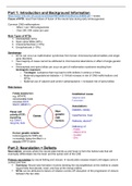
, Process of primary neurulation
1. Closure 1: embryonic day 8.5 at level of hindbran/cervical boundary failure =
craniorachischisis (Copp et al., 2003)
2. Closure 2: initiates at second site on embryonic day 9 in the caudal forebrain or forebrain/midbrain
boundary
a. Initial contact + fusion established between tips of neural folds
b. Failure = anencephaly (cranial NTD)
i. Incomplete ‘zippering’ between closures 1 and 2 which closes the midbrain and
hindbrain
ii. If fusion does not progress from anterior end of neural plate (closure 3), then the
phenotype is split face + forebrain anencephaly
3. Closure 3: Closure spreads bidirectionally in a caudal direction from rostral end
a. Open regions of neural folds (neuropores) shorten and lead to complete closure of the
anterior neuropore on embryonic day 9 and the hindbrain neuropore later
4. Final closure: day 10 spinal neurulation is entirely caudally directed as embryo continues to grow
a. Failure = spina bifida size of ensuing lesions is directly related to the axial level at which
the closure stops
Secondary neurulation by canalisation: tube forms by hollowing out the interior of a solid precursor
1. Defect – lower spinal cord is tethered to the surrounding tissue
2. Solid cord of NT progenitor cells in the developing tail bud becomes canalised to form a
neuroepithelium surrounding a lumen without formation or closure of neural folds (Copp et al., 2015)
3. Involves condensation of tail bud-derived cells to form an epithelial rod that undergoes canalisation
to form the lumen of the tube
4. Malformations = closed NTDs and often involve tethering of the SC with associated ectopic
lipomatous material
Part 3: Mechanisms Underlying Neural Tube Closure
Argument: convergent extension initiates closure
Concomitant with the onset of neural tube closure, the neural plate undergoes narrowing in the mediolateral
axis (convergence) and elongation in the rostrocaudal axis (extension), owing to intercalation of cells at the
midline (Keller 2002).
https://www.ncbi.nlm.nih.gov/pmc/articles/PMC4486472/pdf/emss-63689.pdf = review
Cause of NTD: result from failure of fusion of the neural tube during early embryogenesis
Common CNS malformations
- Affect 1 per 1000 pregnancies
- Over 250, 000 cases per year
Main Types of NTDs
1. Anencephaly (40%)
2. Open spina bifida (40%)
3. Craniorachischisis (<10%)
4. Encephalocele (~10%)
Syndromes
NTDs can occur as malformation syndromes from known chromosomal abnormalities and single
gene disorders
Vast majority of cases cannot be attributed to chromosomal aberrations or effect of single genetic
locus
Anencephaly and spina bifida can occur as part of malformation syndrome resulting from
teratogenic exposure
o Teratogen: substance that may lead to birth defects in embryo or fetus
o Maternal pregestational diabetes = 2-10-fold increase in risk of CNS malformations and
NTDs
o Maternal use of valporid acid and/or carbamazepine = increased risk of spina bidida
Risk factors
Part 2: Neurulation + Defects
Neurulation: process where the neural plate bends up and fuses to form the hollow tube that will
eventually differentiate into the brain and the spinal cord of the CNS
Primary neurulation by neural folding and closure neural plate creases inward until edges come in
contact and fuse
Summary: Neural tube formation involves bending the neuroepithelium at the midline to create
neural folds that elevate, meet, and fuse at the dorsal midline
NTDs can be attributed to failure of initiation events OR disruption of the progression of closure
between the two sites
, Process of primary neurulation
1. Closure 1: embryonic day 8.5 at level of hindbran/cervical boundary failure =
craniorachischisis (Copp et al., 2003)
2. Closure 2: initiates at second site on embryonic day 9 in the caudal forebrain or forebrain/midbrain
boundary
a. Initial contact + fusion established between tips of neural folds
b. Failure = anencephaly (cranial NTD)
i. Incomplete ‘zippering’ between closures 1 and 2 which closes the midbrain and
hindbrain
ii. If fusion does not progress from anterior end of neural plate (closure 3), then the
phenotype is split face + forebrain anencephaly
3. Closure 3: Closure spreads bidirectionally in a caudal direction from rostral end
a. Open regions of neural folds (neuropores) shorten and lead to complete closure of the
anterior neuropore on embryonic day 9 and the hindbrain neuropore later
4. Final closure: day 10 spinal neurulation is entirely caudally directed as embryo continues to grow
a. Failure = spina bifida size of ensuing lesions is directly related to the axial level at which
the closure stops
Secondary neurulation by canalisation: tube forms by hollowing out the interior of a solid precursor
1. Defect – lower spinal cord is tethered to the surrounding tissue
2. Solid cord of NT progenitor cells in the developing tail bud becomes canalised to form a
neuroepithelium surrounding a lumen without formation or closure of neural folds (Copp et al., 2015)
3. Involves condensation of tail bud-derived cells to form an epithelial rod that undergoes canalisation
to form the lumen of the tube
4. Malformations = closed NTDs and often involve tethering of the SC with associated ectopic
lipomatous material
Part 3: Mechanisms Underlying Neural Tube Closure
Argument: convergent extension initiates closure
Concomitant with the onset of neural tube closure, the neural plate undergoes narrowing in the mediolateral
axis (convergence) and elongation in the rostrocaudal axis (extension), owing to intercalation of cells at the
midline (Keller 2002).