Systems
1 THE DIGESTIVE SYSTEM
1.1 THE LAYOUT & STRUCTURE OF THE DIGESTIVE SYSTEM
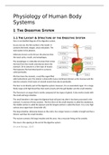
Here is my labelled diagram of the digestive system.
As you can see, the first section is the mouth. It
contains the teeth, tongue, uvula and palate. The
next section is the pharynx.
Otherwise known as the throat, the pharynx links
the nasal cavity, mouth, and oesophagus.
The oesophagus is a tube-like structure that carries
the food from the mouth and pharynx down the
stomach. On its exterior is a thin layer of muscle
that squeezes the food downward in a process
called peristalsis.
We then have the stomach, a sack-like organ filled
with hydrochloric acid. The interior is lined with mucus membrane (known as the mucosa) and the
exterior contains 3 thin layers of smooth muscle that also do peristalsis.
The liver is not directly part of the digestive system; however, it is an associated organ. It is a large,
fleshy organ with high blood flow that works closely with the gall bladder and the small intestine.
The Pancreas is an organ that is mainly composed of two types of glands. It also works closely with
the small and large intestine.
The small intestine is the organ the digested food will pass into after it has been processed in the
stomach. It consists of three sections. The first 20cm of the small intestine is called the duodenum,
the middle section is called the jejunum and the longest section is called the ileum. It is a very high
surface area organ that is lined with villi.
The large intestine, otherwise known as the colon, has a larger lumen, is smaller in length and has a
thinner wall than the small intestine.
The rectum connects the large intestine and the anus. Has a muscular lining on the outside.
The anus is the opening at the end of the digestive system.
(A-Level Biology, 2017)
,1.2 THE FUNCTIONS IN THE DIGESTIVE SYSTEM
I will now talk about each organ in greater detail and explain its function and links within the
digestive system.
1.2.1 The Mouth
At the beginning of the digestive system is the mouth.
When food enters, teeth will be used to grind up the food into smaller chunks. While chewing, the
tongue will push the food around your mouth to ensure it gets thoroughly masticated (chewed). This
is mechanical digestion. Your mouth will also release saliva while eating. Saliva consist of three main
components. Mucin, which binds the food together into ball like clumps (called a bolus) so that it
passes through the oesophagus easier, salivary amylase which starts breaking down starch into
maltose, and mineral salts that will regulate the pH of the food at about 7. This is Chemical digestion.
Once fully masticated, the tongue will push the food backwards, towards the oesophagus.
1.2.2 The Oesophagus
Once the food has passed into the oesophagus, it will get pushed downwards by waves of muscular
movements that squeeze the bolus produced by the mouth into the stomach. This muscle
movement is called peristalsis and is a feature that digestive organs commonly use to move food
around.
At the top of the oesophagus, the peristalsis is voluntary meaning that you can decide if you want to
swallow the food or not. Further down it becomes involuntary and the food will be moved
regardless. It takes between 4-10 seconds for the food to travel the Oesophagus.
1.2.3 The Stomach
(Taylor, 2020) (A-Level Biology, 2017)
When the bolus enters the stomach, the oesophageal sphincter located at the end of the
oesophagus will close behind it, stopping it from being squeezed out later.
In the stomach is a mixture known as gastric juice It consist of Hydrochloric Acid, Mucus, and
digestive enzymes. Its pH varies between 1.5 and 3.5 as this is the optimal pH for enzymes to
function.
Both mechanical and chemical digestion occur. The stomach is externally lined with three layers of
smooth fibre muscle arranged with their fibres running in 3 different directions. This enables it to
contract and expand, thus churning the food inside and allowing it to push food out once it has been
processed. This is peristalsis.
The chemical digestion occurs at the same time as the mechanical digestion. The gastric lipase splits
triglyceride fats into fatty acids and diglycerides and the pepsin breaks proteins into smaller amino
acids. Note that this is not actually full digestion, it is mostly just the stomach preparing some of the
harder-to-digest molecules for further break down by the small intestine. This mix of food and
gastric juice is called chyme.
Usually the stomach is comfortable with holding 1-2 litres of food at once however, it can reach 3-4
litres if needed (if the subject has overeaten or had a large meal). When it gets this full, the stomach
will struggle to digest the food because it will have less room to move during peristalsis. Food usually
remains in the stomach for approximately 1-2 hours.
, Once all the food has been turned to chyme of the correct consistency and pH, the pyloric
sphincter will open at the opposite end of the stomach and release a small amount of the semi
digested food into the duodenum. This release of the chyme will occur over 1-3 hours to keep the
rate of digestion for the small intestine constant and efficient.
1.2.4 The Liver & Gall Bladder
(Taylor, 2020)
The liver plays an important role in digestion because it is the organ that produces bile. Bile is
essentially a mix of water, bile salts, cholesterol and a pigment called bilirubin. Cells called
hepatocytes make the bile which is then passed via bile ducts into the gall bladder where it gets
stored.
When fatty foods enter the duodenum, it will send a hormone signal to the gall bladder to release
bile. Once the requested bile has arrived in the duodenum, it will start to emulsify fats. This means
that the large clumps of fat will be broken up into much smaller pieces, thus increasing the surface
area and making it significantly easier for the rest of the intestine to digest.
1.2.5 The Pancreas
(Taylor, 2020)
The pancreas is essentially two glands. An exocrine gland and an endocrine gland. For digestion, we
will look at its function as an exocrine gland.
As the chyme slowly gets released into the duodenum, the exocrine portion of the pancreas will
release pancreatic juice. Pancreatic juice is a mixture of water, salts, bicarbonate, and many different
digestive enzymes that will be used to complete the chemical digestion that was started by the
stomach. The bicarbonate will neutralise the acidic chyme to protect the walls of the intestine.
Here is a basic explanation of each of the pancreatic enzymes. I will go into this in further detail
later:
Pancreatic Amylase – Breaks down polysaccharides like glycogen and starches into small
sugars that the intestine can absorb such as maltose, maltotriose, and glucose.
Trypsin, Chymotrypsin, and Carboxypeptidase – Protein specific enzymes that break proteins
into amino acids that can be absorbed
Ribonuclease and Deoxyribonuclease – Enzymes that break down nucleic acids like DNA and
RNA into absorbable nutrients.
Pancreatic Lipase – An enzyme that breaks the already semi-broken-down
triglyceride molecules into fatty acids and monoglycerides. These are then emulsified by the
bile release by the gall bladder for further digestion
1.2.6 Small Intestine
1.2.6.1Duodenum
The first section of the small intestine and is about 20cm in length. Like the stomach, it also has a
layer of mucus that serves to protect it from the acidic chyme.
The duodenum is the location where the final stages of digestion start such as the emulsification of
fats via bile, and the release of pancreatic juices.
The duodenum also uses peristalsis to churn the chyme, bile, and pancreatic juice mixture and to
keep the mixture moving along the intestinal tract.

