BHCS2018 Summary Notes
Cardiovascular system (CVS)
Pressure in the cardiovascular system (CVS)
• Fluctuates in ventrioles (0-115mmHg)
• Peak systolic pressure reached in aorta (120mmHG)
• Arterioles are resistance vessels – large drop in pressure
o Contract and dilate to change resistance and regulate flow
o Disorder of these vessels causes clinical hypertension
• Venous pressure is very low (5-10mmHg)
• Capillaries are tiny but numerous
o Total accumulative cross-sectional area is huge
o Velocity = flow/area therefore, increased area, decreases velocity (if flow is constant)
o Decreases velocity allows for effective material exchange in capillaries
• Oscillating pressure is systolic diastolic pressure
• Easier to collect average (mean) of arterial pressure rather than oscillating pressure – gives mean arterial pressure
(MAP)
• Central arterial pressure (pressure in aorta) is approx. halfway between systolic and diastolic pressure
• Pulse pressure = systolic – diastolic
• MAP = diastolic + 1/3 pulse pressure
• Factors of arterial pressure
o Pulse pressure – pressure rises during each pulse
o Stroke volume ejected by l.ventricle into aorta – bigger SV, bigger pulse pressure (i.e. bigger pressure rise)
o Compliance of elastic arteries – greater compliance, easier SV is accommodated in aorta so less pressure
increase
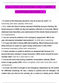
• Arterial compliance curve
o Shows relation between arterial pressure and volume of blood in elastic arteries
o Shows how addition of a given stroke volume creates a certain pulse pressure
o Not linear
o Artery is stiff (low compliance) at higher pressures
• Factors affecting MAP
o Cardiac output (CO = SV * HR)
o Total peripheral resistance (TPR)
• Measuring blood pressure
o Blood pressure taken from upper arm – gives branchial artery pressure (closer to diastolic pressure)
o Arm cuff cuts blood flow at upper arm and is slowly released
o Pressure at which first noise is heard is systolic pressure
o Pressure at which noise disappears is diastolic
Cardiac cycle
• Describes events during one heartbeat
• Mechanical events
o Ventricular diastole is two thirds of cycle
o As ventricle contracts, apex of heart taps against chest wall – used as clinical sign of heart failure as location
of tapping moves during enlargement
o Longitudinal shortening of l. ventricle during systole accounts for 60% stroke volume
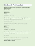
• Ventricular volume changes
o Most filling occurs during early diastole
o Isovolumetric – volume says the same while pressure increases
1
,BHCS2018 Summary Notes
o End-diastolic volume (EDV) – max amount of blood in ventricle (120ml)
o Residual volume – ventricle is never completely emptied, 40ml resides
o Stroke volume – max blood ejected by l. ventricle, EDV – ESV usually 70-80ml
o Ejection fraction – SV/EDV (*100) usually a %, normally 50-70%, declines during heart failure
• Pressure changes
o Mitral valve closed when ventricle exceeds atrial pressure at start of ventricle systole
o Steep increase during isovolumetric contraction in l. ventricle – max rate of rise (dP/dtmax) used as index of
cardiac health
o As l. ventricle – pressure exceeds aortic, aortic valve pushed open to begin ejection (increases arterial blood
pressure)
o As l. ventricle pressure drops, blood flow slowed until slight backflow closes aortic valve – creates slight
notch in aortic pressure trace (incisura)
o Aortic pressure slowly falls away as blood flows in peripheral vessels
o Pressure in ventricle falls rapidly during isovolumetric relaxation
o When ventricle pressure falls below atrial pressure, atrioventricular valves open and passive filling begins
o Atrial pressure
Increases during atrial systole
Increases by bulging of atrioventricular valve into atria (end of isovolumetric contraction)
Decrease for atrial relaxation
Increase for passively filling of atria by returning venous blood during ventricular systole
Sharp decrease due to sudden dumping of blood ventricle when atrioventricular valves open
Heart sounds
• Two sounds - lubb-dubb then a pause
• 1st sound (‘lubb’) – closing of the mitral and tricuspid valves at the start of ventricular systole
• 2nd sound (‘dubb’) – closing of the aortic and pulmonary valves at end of ventricle systole
• As l. ventricle systole ends slightly before the right (due to high peripheral resistance), the second sound is
sometimes audibly split
o Sounds like ‘ter-up’
o Exaggerated by inspiration which lowers intrathoracic pressure – this increases right ventricle filling
therefore prolonging r. ventricle ejection
Central venous pressure (CVP)
• Blood flows from peripheral veins to central veins
• CVP usually 0-7mmHg
• As there are no valves between central veins and right atrium, central veins pulsate
• Assessed by jugular vein in neck
• Raised CVP and jugular filling indicate heart failure
• CVP = V / compliance
• Controlling CVP - Filling pressure of RHS of heart is the CVP - therefore factors affecting CVP will affect stroke
volume
o Volume of blood in circulation
o Distribution of blood between central and peripheral veins
o Sympathetic never activity – regulates peripheral venous tones
o Gravity – standing up ‘pools’ the venous blood in the legs, reducing CVP and SV
Movement operates calf muscle pump raising CVP and SV
o Venous return DOES NOT control cardiac output – it is cardiac output (as it is the flow of blood through
circulation)
Cardiac control
• Cardiac output (CO = SV * HR)
• Factors affecting stroke volume
o Energy of ventricular contraction
Ventricular filling pressure (Starling’s law)
Myocardial contractility
• Regulated by sympathetic nerves and circulating agents – e.g., adrenaline
2
, BHCS2018 Summary Notes
• A rise in filling pressure or contractility increases energy, therefore increases SV
o Aortic blood pressure
Opposes ventricular ejection
Rise in aortic pressure (due to rise to TPR) causes ventricle to use more energy to reach higher
pressure to open valve – SV decreases
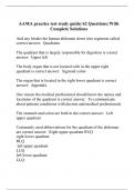
Starling’s law of the heart
• Relationship between stroke volume and end-diastolic volume
• Stroke volume increases with increasingly filling pressure
• Therefore, increasing venous return increases filling pressure which increases SV (so CO increases)
• When l. ventricle end-diastolic pressure reaches 10mmHg or higher, there is no further gain in SV with distension
(plateau)
Laplace’s law
• Relates wall tension to internal pressure
• Pressure = 2 * wall tension / radius
• If radius dilates excessively, and tension plateaus, then systolic pressure generated by contraction will fall SV
decreases
• The bigger radius, the less internal pressure a given wall tension creates
• To improve co, reduce distension of a dilated heart (i.e., decrease dilation)
Balancing the left and right stroke volume
• Filling pressure of the LHS is mainly determined by output of RHS, so therefore CVP (RHS = CVP)
• Starling’s Law ensures the right and left SV are equal over any time intervals longer than a few beats – prolonged
imbalance would have bad consequences for blood volume in pulmonary circulation
• CVP R. atrial pressure R. ventricle EDP R. ventricle EDV R. ventricle ED fibre length R. ventricle
contraction energy R. ventricle SV Pulmonary pressure and volume L. atrial pressure L. ventricle EDP
L. ventricle EDV L. ventricle ED fibre length L. ventricle contraction energy L. ventricle SV
• Starling’s law
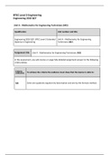
The pressure-volume loop
3
Cardiovascular system (CVS)
Pressure in the cardiovascular system (CVS)
• Fluctuates in ventrioles (0-115mmHg)
• Peak systolic pressure reached in aorta (120mmHG)
• Arterioles are resistance vessels – large drop in pressure
o Contract and dilate to change resistance and regulate flow
o Disorder of these vessels causes clinical hypertension
• Venous pressure is very low (5-10mmHg)
• Capillaries are tiny but numerous
o Total accumulative cross-sectional area is huge
o Velocity = flow/area therefore, increased area, decreases velocity (if flow is constant)
o Decreases velocity allows for effective material exchange in capillaries
• Oscillating pressure is systolic diastolic pressure
• Easier to collect average (mean) of arterial pressure rather than oscillating pressure – gives mean arterial pressure
(MAP)
• Central arterial pressure (pressure in aorta) is approx. halfway between systolic and diastolic pressure
• Pulse pressure = systolic – diastolic
• MAP = diastolic + 1/3 pulse pressure
• Factors of arterial pressure
o Pulse pressure – pressure rises during each pulse
o Stroke volume ejected by l.ventricle into aorta – bigger SV, bigger pulse pressure (i.e. bigger pressure rise)
o Compliance of elastic arteries – greater compliance, easier SV is accommodated in aorta so less pressure
increase
• Arterial compliance curve
o Shows relation between arterial pressure and volume of blood in elastic arteries
o Shows how addition of a given stroke volume creates a certain pulse pressure
o Not linear
o Artery is stiff (low compliance) at higher pressures
• Factors affecting MAP
o Cardiac output (CO = SV * HR)
o Total peripheral resistance (TPR)
• Measuring blood pressure
o Blood pressure taken from upper arm – gives branchial artery pressure (closer to diastolic pressure)
o Arm cuff cuts blood flow at upper arm and is slowly released
o Pressure at which first noise is heard is systolic pressure
o Pressure at which noise disappears is diastolic
Cardiac cycle
• Describes events during one heartbeat
• Mechanical events
o Ventricular diastole is two thirds of cycle
o As ventricle contracts, apex of heart taps against chest wall – used as clinical sign of heart failure as location
of tapping moves during enlargement
o Longitudinal shortening of l. ventricle during systole accounts for 60% stroke volume
• Ventricular volume changes
o Most filling occurs during early diastole
o Isovolumetric – volume says the same while pressure increases
1
,BHCS2018 Summary Notes
o End-diastolic volume (EDV) – max amount of blood in ventricle (120ml)
o Residual volume – ventricle is never completely emptied, 40ml resides
o Stroke volume – max blood ejected by l. ventricle, EDV – ESV usually 70-80ml
o Ejection fraction – SV/EDV (*100) usually a %, normally 50-70%, declines during heart failure
• Pressure changes
o Mitral valve closed when ventricle exceeds atrial pressure at start of ventricle systole
o Steep increase during isovolumetric contraction in l. ventricle – max rate of rise (dP/dtmax) used as index of
cardiac health
o As l. ventricle – pressure exceeds aortic, aortic valve pushed open to begin ejection (increases arterial blood
pressure)
o As l. ventricle pressure drops, blood flow slowed until slight backflow closes aortic valve – creates slight
notch in aortic pressure trace (incisura)
o Aortic pressure slowly falls away as blood flows in peripheral vessels
o Pressure in ventricle falls rapidly during isovolumetric relaxation
o When ventricle pressure falls below atrial pressure, atrioventricular valves open and passive filling begins
o Atrial pressure
Increases during atrial systole
Increases by bulging of atrioventricular valve into atria (end of isovolumetric contraction)
Decrease for atrial relaxation
Increase for passively filling of atria by returning venous blood during ventricular systole
Sharp decrease due to sudden dumping of blood ventricle when atrioventricular valves open
Heart sounds
• Two sounds - lubb-dubb then a pause
• 1st sound (‘lubb’) – closing of the mitral and tricuspid valves at the start of ventricular systole
• 2nd sound (‘dubb’) – closing of the aortic and pulmonary valves at end of ventricle systole
• As l. ventricle systole ends slightly before the right (due to high peripheral resistance), the second sound is
sometimes audibly split
o Sounds like ‘ter-up’
o Exaggerated by inspiration which lowers intrathoracic pressure – this increases right ventricle filling
therefore prolonging r. ventricle ejection
Central venous pressure (CVP)
• Blood flows from peripheral veins to central veins
• CVP usually 0-7mmHg
• As there are no valves between central veins and right atrium, central veins pulsate
• Assessed by jugular vein in neck
• Raised CVP and jugular filling indicate heart failure
• CVP = V / compliance
• Controlling CVP - Filling pressure of RHS of heart is the CVP - therefore factors affecting CVP will affect stroke
volume
o Volume of blood in circulation
o Distribution of blood between central and peripheral veins
o Sympathetic never activity – regulates peripheral venous tones
o Gravity – standing up ‘pools’ the venous blood in the legs, reducing CVP and SV
Movement operates calf muscle pump raising CVP and SV
o Venous return DOES NOT control cardiac output – it is cardiac output (as it is the flow of blood through
circulation)
Cardiac control
• Cardiac output (CO = SV * HR)
• Factors affecting stroke volume
o Energy of ventricular contraction
Ventricular filling pressure (Starling’s law)
Myocardial contractility
• Regulated by sympathetic nerves and circulating agents – e.g., adrenaline
2
, BHCS2018 Summary Notes
• A rise in filling pressure or contractility increases energy, therefore increases SV
o Aortic blood pressure
Opposes ventricular ejection
Rise in aortic pressure (due to rise to TPR) causes ventricle to use more energy to reach higher
pressure to open valve – SV decreases
Starling’s law of the heart
• Relationship between stroke volume and end-diastolic volume
• Stroke volume increases with increasingly filling pressure
• Therefore, increasing venous return increases filling pressure which increases SV (so CO increases)
• When l. ventricle end-diastolic pressure reaches 10mmHg or higher, there is no further gain in SV with distension
(plateau)
Laplace’s law
• Relates wall tension to internal pressure
• Pressure = 2 * wall tension / radius
• If radius dilates excessively, and tension plateaus, then systolic pressure generated by contraction will fall SV
decreases
• The bigger radius, the less internal pressure a given wall tension creates
• To improve co, reduce distension of a dilated heart (i.e., decrease dilation)
Balancing the left and right stroke volume
• Filling pressure of the LHS is mainly determined by output of RHS, so therefore CVP (RHS = CVP)
• Starling’s Law ensures the right and left SV are equal over any time intervals longer than a few beats – prolonged
imbalance would have bad consequences for blood volume in pulmonary circulation
• CVP R. atrial pressure R. ventricle EDP R. ventricle EDV R. ventricle ED fibre length R. ventricle
contraction energy R. ventricle SV Pulmonary pressure and volume L. atrial pressure L. ventricle EDP
L. ventricle EDV L. ventricle ED fibre length L. ventricle contraction energy L. ventricle SV
• Starling’s law
The pressure-volume loop
3