100% satisfaction guarantee Immediately available after payment Both online and in PDF No strings attached
logo-home
Summary OCR Biology F211 WORD DOCUMENT £3.49
Add to cart

Summary

Summary OCR Biology F211 WORD DOCUMENT

 1 purchase

Notes made based on: CGP AS and A2 OCR Biology textbook OCR AS Biology Student Book OCR Specification Basically a condensed version of all of these cutting out the BS that's not needed; learn these by heart and do some past papers and that grade A/B is yours. I used these for the July 2015 Biolo...

[Show more]
Preview 6 out of 27  pages

  • July 19, 2015
  • 27
  • 2014/2015
  • Summary
  • biology
  • ocr
  • college
  • 2015
  • 2014
  • 2016
  • aqa
All documents for this subject (1)
avatar-seller
sal98
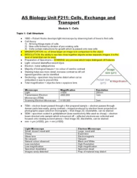
AS Biology Unit F211: Cells, Exchange and
Transport
Module 1: Cells
Topic 1: Cell Structure

● 1660 – Robert Hooke develops light microscope by observing bark of trees to find cells
● Cell theory:
1) All living things made of cells
2) New cells formed by division of pre-existing cells
3) Cells contain instructions for growth which is passed onto new cells
● MAGNIFICATION: no. of times larger an image is in comparison to the object
● RESOLUTION: the ability to see two close together objects as two separate images; it is the
level of detail that can be seen
● Preparation of Specimens – STAINING: any process which helps distinguish dif features
● Light: coloured stains/fluorescent dyes
● Electron: metal salts/particles
● Majority of biological tissues = no colour of own/no contrast
● Staining helps see more detail, increase contrast so dif cell
types/organelles can be identified
● Sectioning – specimen may become distort when cut so
embedded in wax to prevent this
● Total magnification = objective lens x eyepiece lens


Microscope Magnification Resolution
Light x1500 200nm
Transmission Electron x500,000 1nm
Microscope (TEM)
Scanning Electron Microscope X100,000 2nm

● TEM – electron beam passed through v thin prepared sample – electron passes through
denser parts less easily, giving contrast – imaged produced by electron beam projected on
photographic paper (electron micrograph) – final image 2D, black/white, can be stained
● SEM – specimen coated in gold/platinum (less reactive/only little used so cheap) – electron
beam directed onto sample which is bounced off – reflected electrons are collected and
focused onto viewing screen/camera – final image 3D, black/white, can be stained
● mm -> µm (x1000), µm -> nm (x1000)


Light Microscope Electron Microscope
Small, doesn’t need skill Large, need skill
Cheap Expensive
Glass lens Electromagnets
No vacuum Vacuum
Natural colour observed Black and White
Low resolution High Resolution
Low magnification High magnification
Specimen dead/alive Specimen dead
Images rarely distorted Prep of material may distort

, Organelle Structure Function
Nucleus and -Nucleus: largest organelle -Nucleus: contains DNA
Nucleolus -Nucleolus: dense, spherical -Nucleolus: role in mRNA,
religion in nucleus ribosome synthesis
Rough Endoplasmic -Flattened membrane-bound -Transports proteins around cell
Reticulum (RER) sacs (cisternae)
-Studded w/ ribosomes
Smooth Endoplasmic -Flattened membrane-bound -Lipid synthesis
Reticulum (SER) sacs (cisternae)
-No ribosomes
Golgi Apparatus -Stack of membrane-bound sacs -Modifies and packages proteins
(cisternae)
Ribosomes -Smallest organelles -Protein synthesis
-Spherical, 2 sub-units
-Free bound or bound to RER
-Not membrane bound
Mitochondria -Spherical/sausage-shaped -ATP synthesis for aerobic resp.
-2 membranes
Lysosomes -Spherical, surrounded by single -Has digestive enzymes to break
membrane down waste material
Chloroplasts / -Light for photosyn.
Centrioles -Form rings outside nucleus -Spindle fibres grow out of them
-Only in animal cells during cell division
Cytoplasm -Jelly-like substance -Where reactions take place
● Protein Synthesis:
1) mRNA makes copy of the insulin (protein) gene in the nucleus
2) mRNA leaves via nuclear pores
3) mRNA attaches to ribosome (free-bound or RER) which uses code to produce protein
(hormone)
4) Transport vesicles bud off from RER -> golgi apparatus
5) Vesicles fuse w/ golgi
6) Hormone process, modified and packaged for release
7) Vesicles bud off from golgi -> cell surface membrane
8) Secretory vesicles fuse w/ cell surface membrane and contents are exported by
exocytosis
● ORGANELLE: particular structure of a cell that has a specialised function
● ULTRASTRUCTURE: detail of the inside of the cell as seen w/ an electron microscope
● DIVISION OF LABOUR: each type of organelles has a specific role within a cell where they
work together and contribute to cell survival
● Cells contain network of fine filaments/fibres that make up cytoskeleton; actin filaments,
microtubules (made of protein tubulin)
● Cytoskeleton involved in:
Providing cell support/strength
Cell division (centrioles & spindle fibres)
● Movement of:
-Chromosomes –Secretory vesicles –mRNA (in protein syn) –holding/moving organelles
-cytoplasm in cytokinesis –cytoplasmic streaming
● Microtubules = key components of cilia, flagella and centrioles
● Cilia: numerous, short, hair like ext. sticking out of cell surfaces, move in wave-like pattern
e.g. windpipe
● Flagella: long, fewer in no., move whole cell e.g. sperm
● VESICLES: membrane-bound sacs that carry substances around cells

, ● LARGE CONTRACTILE VACUOLE: filled w/ water and solutes which maintain cell’s
stability/turgidity
● PLANT CELL WALL: made up of cellulose which provides strength/support to plants
● EUKARYOTIC: organisms that have cells w/ a membrane-bound nucleus, ‘true nucleus’
● PROKARYOTIC: organisms that lack membrane-bound nucleus

Eukaryotic Prokaryotic
Nucleus No nucleus
Linear DNA Circular DNA
DNA associated w/ histone proteins DNA not associated w/ histone proteins
Membrane-bound organelles No membrane-bound organelles
May have cell wall (cellulose) Cell wall (peptidoglycan)
Larger 80s ribosomes Smaller 70s ribosomes
No plasmids (except inside organelles) Plasmids
Cytoskeleton No cytoskeleton
Flagellum 9+2 Don’t have 9+2
No pili/capsule/mesosome Pili/capsule/mesosome
● Prokaryotes – diseases, some bacterial strains are resistant to antibiotics, resistance coded
on plasmids and passed between cells etc.
● Good uses – food industry, mammalian intestines, normal flora, sewage system

Topic 2: Cell Membranes

● Phospholipid bilayer = phosphate group w/ fatty acid tails (lipid) w/ proteins embedded
● Bilayer = 2 layers
● Basic structural component of all cells membranes
● 7nm thick
● Roles OUTSIDE cell:
1) Controls what goes in/out – allows lipid soluble substances through but not water soluble
substances
2) Cell recognition and cell signalling
3) Acts as barrier – separates outside environment from contents of the cell (so organelles
don’t spill out)
● Roles INSIDE cell:
1) Compartmentalisation – separates cell organelles from cytoplasm
2) Isolates – isolates harmful substances from organelles e.g. lysosomes
3) Selective permeability – controls what can enter and leave cell
4) Provides surface of attachment – by holding components of some metabolic pathways in
place e.g. ribosomes on RER
● Phospholipid head = hydrophilic, tail = hydrophobic, due to distribution of charges across a
molecule; molecules w/ charges distributed evenly around them repel water (oil and water)
● Monolayer – phospholipid put in water; head in water, tails stick out
● Micelle – phospholipid mixed w/ water; head faces out, tails towards each other to prevent
interaction
● Bilayer – phospholipid completely surrounded by water; head out, tails towards each other
● Simple phospholipid bilayer:
-can’t perform all biological functions
-too fragile
-other components needed to function
-no. and type of components needed depend on function of the particular membrane (this
specialisation is a part of differentiation)
● Examples:
-Growing shoots: receptors to detect molecules that regulate growth
-Muscle cells: contain numerous channels for glucose uptake
-Chloroplasts: molecules for photosynthesis
-White Blood Cells: cell recognition proteins

,● Permeability: partially permeable membrane allows for selective substances to pass, all
membranes are permeable to water but some have aquaporin (protein channels that allow
water molecules through) which are more permeable to water

● Fluid Mosaic Model:
-used to describe the molecular arrangement in the
membrane
-phospholipid molecules form a continuous,
double layer
-bilayer is ‘fluid’ as phospholipids are always
moving
-cholesterol molecules between tails
-proteins scattered throughout hence mosaic
-some proteins have polysaccharide attached:
glycoproteins
-some lipids have polysaccharide attached:
glycolipid
● Membranes and temp.:
-increase in temp. = more kinetic energy
-move faster
-increased movement of phospholipids and other components makes membranes leaky
-ends up allowing substances through that aren’t meant to go through
-proteins denature
● Cell signalling:
-communication between cells using signals; molecule is released by one cell, attaches to or
causes change in another cell
-this is important between multicellular organisms as it
1. Regulates development and organisation
2. Controls growth and cell division
3. Co-ordinates various cell activities
● Signals: many molecules can act as signals e.g. cytokines, drugs, neurotransmitters etc.
● Water soluble: cannot pass through membrane
● Water insoluble: can pass through membrane
● Receptors: protein molecules on the cell surface that act as ‘sensors’ which are capable of
receiving signals; these are specific and complimentary to the signal
● Hormones: chemical messenger produced in specific tissues and released directly into the
bloodstream
● Target cell: any cell w/ a receptor for the hormone molecule
● Cell membrane and cell signalling
1. Signal molecule is released by exocytosis/secretion in a specific tissue
2. Proteins/glycolipids/proteins act as receptors
3. Shape of receptor and signal are specific and complimentary to one another
4. Signal molecules attaches to the receptor of the target cell and causes a change
(inside cell/cell surface)
5. Cell surface membrane only allows for some signal molecules to enter
● Medicinal drugs:
-numerous drugs are complimentary to the shapes of specific receptor molecules e.g. beta
blockers (lowers heart pressure by blocking the receptors)
● Hijacking receptors:
-viruses bind onto human receptors
-HIV binds onto T-cells and enters into it where it reproduces and destroys the cell
● Botox:
-botulinum toxin: binds to Acetylcholine receptors on muscle cells
-block the receptors so that the neurotransmitter Acetylcholine cannot bind to it during muscle
contraction
-causes paralysis

, Membrane Component Description Function
Phospholipid Bilayer made of glycerol, -Forms basic structure of
phosphate and fatty acid tails membrane
w/ proteins embedded w/i -small, non-polar substances
them diffuse through
Glycoprotein Protein w/ carbohydrate -Acts as antigens (cell
attached surface membrane involved
in the immune response)
-Act as receptors (for cell
signalling)
-Receptor sites (for
hormones, antibodies and
drugs)
-Attach to water molecules
(to stabilise the membrane)
Glycolipid Lipid w/ carbohydrate ‘’
attached
Cholesterol Steroid molecule that fit -Mechanical stability
between fatty acid tails
Channel Protein Intrinsic protein that spans the -Movement of polar
bilayer substances across
membrane
Carrier Protein ‘’ -Movement of large polar
substances across
membrane
Receptor Sites These may be -Hormones bind to specific
glycoproteins/lipids receptor for a cell response
Enzymes and co-enzymes Free-bound of bound to -Allow for important
cristae (electron transfer reactions to take place
reactions) or inside the
chloroplast thylakoids
(chlorophyll)

● Transport across membranes:
-phospholipid bilayer is an effective barrier to water soluble molecules and ions
-exchange of substances happen by:
1. Diffusion
2. Facilitated Diffusion
3. Osmosis
4. Active Transport
5. Bulk Transport
(first three are passive/don’t need ATP)
● Diffusion: movement of a substance from a region of high conc. to a region of low conc. down
a conc. gradient; eventually equilibrium is reached
● Facilitated Diffusion: movement of a substance from a region of high conc. to a region of low
conc. down the conc. gradient using proteins; eventually equilibrium is reached

, ● Diffusion:
-small molecules e.g. oxygen and carbon dioxide
-lipid-based molecules e.g. steroid hormones
● Factors that affect diffusion rate:
1. Temp. - gives molecules more kinetic energy
2. Conc. gradient - more molecules on one side of the membrane
3. Stirring/moving - increases movement of the particles
4. Surface Area – greater S.A for diffusion to occur
5. Distance/thickness – greater distance for molecules to travel
6. Size – smaller ions/molecules diffuse faster
● Facilitated Diffusion:
Channel protein:
-forms pore through centre of protein, shaped to allow one type of ion through
-hydrophilic conditions
-may be gated
-polar molecules e.g. sodium and calcium ions
Carrier protein:
-specific shape for when the molecule fits in, then changes shape so molecules goes through
to other side (doesn’t need ATP)
-larger polar molecules e.g. glucose and amino acids
● Limitations of diffusion when substances need to move across membrane:
-against conc. gradient
-faster than diffusion allows e.g. Mg ions in soil, absorbing glucose from intestine
● Active Transport:
-movement of substances across a membrane against the conc. gradient which uses ATP and
carrier proteins
-carrier proteins (acting as pumps) are complimentary to shapes of molecules they carry
Different to diffusion:
-dif proteins used to FD (carrier proteins acting as protein pumps)
-use ATP
-carry molecules against conc. gradient
-faster
Ensure one way flow:
-carrier proteins are specific and complimentary to molecules they carry
-fits into one side of membrane
-ATP changes shape of protein
-molecule released to other side of membrane
-can’t re-enter protein as now dif shape
● Bulk Transport:
● Endocytosis – bringing materials into cell
● Exocytosis – moving materials out of the cell
● Endo – inwards
● Exo – outwards
● Phago – solid
● Pino – liquid
● E.g. moving liquid out of cell = exopinocytosis
● Endocytosis:
-membrane folds in, fuses w/ self, pinches off to form vesicle, vesicle moves through
cytoplasm using ATP
● Exocytosis:
-vesicle moves through cytoplasm using ATP, fuses w/ plasma membrane discharging
releasing contents into the exterior
● Movement of membrane to form vesicle + movement of secretory vesicles around cell both
require ATP
● Examples:

The benefits of buying summaries with Stuvia:

Guaranteed quality through customer reviews

Guaranteed quality through customer reviews

Stuvia customers have reviewed more than 700,000 summaries. This how you know that you are buying the best documents.

Quick and easy check-out

Quick and easy check-out

You can quickly pay through credit card for the summaries. There is no membership needed.

Focus on what matters

Focus on what matters

Your fellow students write the study notes themselves, which is why the documents are always reliable and up-to-date. This ensures you quickly get to the core!

Frequently asked questions

What do I get when I buy this document?

You get a PDF, available immediately after your purchase. The purchased document is accessible anytime, anywhere and indefinitely through your profile.

Satisfaction guarantee: how does it work?

Our satisfaction guarantee ensures that you always find a study document that suits you well. You fill out a form, and our customer service team takes care of the rest.

Who am I buying these notes from?

Stuvia is a marketplace, so you are not buying this document from us, but from seller sal98. Stuvia facilitates payment to the seller.

Will I be stuck with a subscription?

No, you only buy these notes for £3.49. You're not tied to anything after your purchase.

Can Stuvia be trusted?

4.6 stars on Google & Trustpilot (+1000 reviews)

62774 documents were sold in the last 30 days

Founded in 2010, the go-to place to buy revision notes and other study material for 15 years now

Start selling
£3.49  1x  sold
  • (0)
Add to cart
Added