Mohammed Salam Unit 8 LA B
Impact of lymphatic disorder and associated treatments
Introduction:
In this written report, I will discuss about the anatomy and functions of the organs in the lymphatic
system, the formation of the lymph, I will go in depth about the disorder in the lymphatic system like
lymphedema and I will also discuss the physiological reasoning for each treatment and a patient case
study.
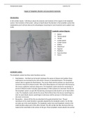
Lymphatic system diagram:
Spleen
Thymus gland
Tonsils
Lymph glands
Lymph vessels
Axillary
Abdominal
Inguinal
Popliteal
Supratrochlear
Valves
Lymphatic system
The lymphatic system has three main functions such as:
Fluid balance – the fluid can be found in between the spaces of tissues and cavities; these
small spaces are enclosed by the cells which is known as interstitial spaces. The lymphatic
system aids to regulate the fluid balance. The excess fluid and the proteins are returned but
cannot be returned via blood vessels. 90% of the blood plasma are returned in the tissues by
the venous capillaries and back along veins. The lymphatic system drains the remaining 10
percent of blood vessels. Everyday approximately 2-3 litres plasma are returned. The loss of
the lymphatic system can give life threatening consequences like death it can be fatal within
a day. The lymphatic system drains the excess fluid without them the tissues would begin to
start to swell, blood volume would begin to decrease and the pressure in the body would
also start to increase.
Absorption – almost all the fats are absorbed in the gastrointestinal tract. The gut
membrane in the small intestine is specially adapted by the lymphatic system. So, the fats
are taken up in the small intestine. The lymphatic system has small lymphatic vessels in the
small intestine where part of the villi is formed. The villi are like hair like structures that are
made by the small folds in the surface of the gut. These lymphatic vessels absorb fats and fat
1
, Mohammed Salam Unit 8 LA B
soluble like vitamins and forms in result of milky white fluid which is called chyle. The white
fluid contains lymph and free fatty acids. When it reaches the venous blood circulation, it
indirectly delivers nutrients. The blood capillaries deliver other nutrients directly.
Defensive mechanism – human body are exposed to deadly pathogens, but the human has
defence in the first line like skin, acidic contents in the stomach and good bacteria in the
body. But these first line of defence can sometimes fail so the lymphatic system are involved
to release the white blood cells to exterminate the deadly pathogens. Number of different
variants of immune cells and special molecules cooperates to fight these pathogens. If the
immune system fails to fight these pathogens it can be harmful for the body and can be
fatal.
Maintenance of hydrostatic pressure:
“Hydrostatic pressure is maintained by the arterioles, the smallest vessels on the arterial side of the
vasculature. Arterioles respond to changes in pressure and/or flow via their myogenic response.”
(Davis & Hill, 1999).
As a result of hydrostatic pressure, lymphatic capillaries takes lymph fluid from the tissues, in order
to maintain the interstitial fluid pressure. When interstitial fluid accumulates, and the pressure in the
lymph exceeds the pressure in the lymph, the small valve opens, allowing the fluid to enter the
lymphatic capillary. Lymphatic capillaries stick closer together when there is a high level of pressure
inside, preventing lymph backflow. When tissues swell, anchoring filaments are also pulled apart. By
doing so, the lymph capillaries expand further, increasing their capacity and decreasing their
pressure. There is a higher hydrostatic pressure in lymph capillaries due to the higher concentration
of proteins in the lymph than blood plasma due to the higher concentration of plasma proteins in
lymph. In addition, lymphatic capillaries are larger than cardiovascular capillaries, allowing them to
take in more fluid proteins into lymph than plasma, contributing to their higher hydrostatic pressure.
Proteins exert hydrostatic pressure, so fluid follows them, and lymph flows easily into lymph
capillaries.
Filtration:
Due to capillary hydrostatic pressure (35 mm Hg) is larger than blood colloidal osmotic pressure,
fluid escapes the capillary at 25 mm Hg.
No net movement:
Since capillary hydrostatic pressure (25 mm Hg) equals blood colloidal osmotic pressure, there is no
net fluid movement (25 mm Hg).
Reabsorption:
Since capillary hydrostatic pressure (18 mm Hg) is less than blood colloidal osmotic pressure, fluid re-
enters the capillary (25 mm Hg).
Spleen:
2


