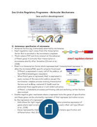
Sea Urchin Regulatory Programme – Molecular Mechanisms
1) Autonomous specification of micromeres
Maternal factors (eg. Disheveled) inherited by micromeres
Next regulatory input comes from Otx transcription
factor that is enriched in the micromere cytoplasm
The β-catenin/TCF and Octx interact at the enhancer
of Pmar1 genes to activate their transcription in
micromeres shortly after formation (Olivieri et al.,
2008)
Pmar1 is a transcription factor which represses hesC transcriptional repressor to
allow the micromere/PMC-specific program to proceed
- If Pmar1 is expressed in every cell of the embryo, all
form PMCS/skeletogenic mesoderm
- When Pmar1 gene is repressed, HesC is expressed in
every nucleus of the sea urchin embryo except the
micromeres: complex process involving transcription
factors such as Blimp, maternal tf SoxB1 must be
eliminated from vegetal pole or it will inhibit activation
of Pmar1, cytoskeletal processes partitioning cells and anchoring certain factors
also involved
Double-negative gate: mechanism where a repressor locks the genes of specification
and these genes can be unlocked by repressor of that repressor (activation occurs
by the repression of a repressor)
- Gate allows for tight regulation of fate specification promotes expression of
genes where input occurs, represses same gene in every other cell type (Oliveri
et al., 2008)
- Genes repressed by HesC are those involved in micromere specification and
differentiation: Alx1, Ets1, Tbr, Tel, SoxC
, Each of these genes can be activated by ubiquitous transcription factors but
positive transcription factors cannot work while HesC repressor protein binds to
the respective enhancers
When Pmar1 protein is present, it represses HesC, all these genes become active
(Revilla-i-Domingo et al., 2007)
- Newly activated genes synthesise transcription factors that activate another
set of genes, most of which are genes that activate skeletal determinants
- Transcription factors also activate each other’s genes, so once one factor is
activated, maintains the activity of other skeletogenic genes – this stabilises
the regulatory state of the skeletogenic mesenchyme cells
Control of the differentiation genes that make sea urchin skeleton operates on a
feedforward process
- Skeletogenetic portion of the micromere GRN
appears due to the recruitment of a “subroutine”
that in most echinoderms is used
- The regulatory state initiated by transient nuclear β-
catenin progressed and maintained – specification
initiated by pmar1/hesC is stabilised by a 3 gene positive
reciprocal regulation (eg. Erg, Hex, Tgif)
- Early and late micromere genes, directly regulate the differentiation genes –
each differentiation gene is regulated by a combination of factors to ensure
they are expressed only in the right cell type (eg. repression of non-skeletogenic
mesodermal (NSM) fate by Alx1, a gene downstream of the pmar1/hesC system;
in absence of Alx1 SMs acquire NSM features)
Cooption of subroutines by a new lineage is one of the ways evolution occurs
- Happens that GRN of micromeres in sea urchin embryos is very different from
that in other echinoderms
- Only in micromeres of sea urchins have the skeletogenic subroutine (which in all
other echinoderms is activated late in development) come under control of genes
that specify cells to the micromere lineage
- Evolutionary events – placing skeletogenic genes Alx1 and Ets1 (necessary for
adult skeletal development) and Tbr (used in later larval skeleton formation)
under regulation of the Pmar1/HesC double negative gate
- This occurred through mutations in the cis-regulatory regions of these genes –
skeletogenic properties that distinguish sea urchin micromeres appear to have
arisen through co-option of a pre-existing skeletogenic regulatory system by the
micromere lineage gene regulatory system
Evolutionary perspective on PMC ingression, migration of skeletogenic cells,
patterning of skeleton and gastrulation
, Analogous and homologous morphogenetic movements studied in other organisms –
with some general similarities across cell types and phyla
Molecular components most often conserved are those involved in cell-cell adhesion
and cytoskeletal dynamics, properties fundamental to all cell types
Regulatory processes however are divergent – partly because they occur in
differentiated tissues, already in an advanced state of specification
By breaking down morphogenetic processes down to smaller units (eg. cell adhesion,
polarity, shape, motility) subroutines of the network that drive each can be
determined sea urchin GRN is a good model to study these subroutines
Specification of the endomesoderm (veg2)
Micromeres also function as a crucial signalling centre,
inducing differentiation of the mesoderm and endoderm – at
least 3 signals that emanate from the micromeres to pattern
this
Transient input leads to lockdown circuitry installation of
intra- and inter- gene feedback circuits
1. Nuclear β-catenin is maintained by Wnts signalling in
veg2 endomesoderm
- As soon as the micromeres form, maternal β-
catenin and Otx activate the Blimp1 gene, whose
produce (in conjunction with more β-catenin)
activates the gene encoding paracrine factor
Wnt8
- Wnt8 is received by micromeres’ own genes for
β-catenin
- As β-catenin activates Blimp1, this sets up a positive feedback loop between
Blimp1 and Wnt8, establishes a source of β-catenin for the nuclei of the
micromeres (Logan et al., 1998 experiments showed this)
2. Early signal (ES) gene still unidentified


