Neural Crest derivatives
The Neural Crest
Derived from ectoderm, but is so important it
is sometimes called the fourth germ layer
(Hall, 2009)
Led to pivotal events jaws, face, skull,
sensory ganglia of vertebrates
Transient structure – adults/later stage
vertebrate embryos cells of neural crest
undergo an epithelial-to-mesenchymal
transition fr om the dorsal neural tube after
which they migrate extensively to generate a
prodigious number of differentiated cell types
Neural crest is a population of multipotent
progenitor cells that can produce tissues such
as
1. Neurons and glial cells of the sensory, sympathetic and parasympathetic nervous
systems
2. Epipnephrine-producing (medulla) cells of the adrenal gland
3. Pigment-containing cells of the epidermis
4. Many skeletal and connective tissue components of the heads
Remains unknown whether most are already restricted to certain fates or are
multipotent
- Bronner-Fraser and Fraser (1988) provided evidence that many individual trunk
neural crest cells are multipotent as they leave the crest: injected fluorescent
dextran molecules into individual chick neural crest cells while cells still above
the neural tube then looked at the fates of cells after migration
Could become sensory neurons, melanocytes, adrenomedullary cells,
- Henion and Weston (1997) also found initial avian trunk neural crest population
was a heterogenous mixture of precursor cells, that nearly half of the cells
emerge from the neural crest are restricted to generate single cell type
- Model: an original multipotent neural crest cell divides and progressively loses
its development, though whether a neural crest multipotent precursor cell can
give rise to other precursor cells (ie. a true stem cell?)
Specification of Neural Crest Cells
, Specification is a multi-step process
1. Location of the neural plate border
- Amphibians border appears to be
specified by intermediate
concentrations of BMPs
- Raven and Kloos (1945) showed that
presumptive notochord could induce
both amphibian neural plate and neural
crest tissue (presumably blocking
nearly all BMPs), somite mesoderm and
lateral plate mesoderm could only
induce the neural crest
- In chick embryos, specification occurs
during gastrulation – where borders
between neural and non-neural
ectoderm are still forming (Basch et
al., 2006)
Here, neural plate inductive signals
(especially BMPs and Wnts) secreted from ventral ectoderm and paraxial
mesoderm interact to specify the boundaries
2. In anterior region, timing of BMP and Wnt signal expression is critical for
discriminating between neural plate,
epidermis, placode and neural crest
tissues
- If both BMP and Wnt signalling are
continuous, fate of ectoderm is
epidermal, if BMP antagonists (eg,
Noggin or FGFs) block BMP signalling,
ectoderm becomes neural
- Neural plate induces in these border
cells a set of transcription factors –
neural plate border specifiers –
Distalless-5, Pax3 and Pax7,
collectively prevent border region from
becoming either neural plate or epidermis
- Border specifying transcription factors then induce second set of tfs – neural
crest specifiers in those cells fated to become neural crest (FoxD3, Sox9, Id,
Twist, Snail)
, - When FoxD3, Snail,, Sox3 experimentally expressed in lateral neural tube, these
lateral neuroepithelial cells become neural crest-like, undergo epithelial-
mesenchymal transition, delaminate from then neuroepithelium
- Sox9 and Snail suffiecient to induce transition
- Sox9 is needed for the survival of trunk neural crest cells after delamination (in
the absence, neural crest cells undergo apoptosis as soon as they delaminate)
- FoxD3 plays many roles: needed for expression of the cell surface proteins
needed for migration, critical for specification of the ectodermal neural crest
- Inhibiting FOxD3 gene inhibits neural crest differentiation
- FoxD3 expressed ectopically by electroporating active gene into neural plate
cells, neural plate cells express proteins characteristic of the neural crest
(Cheung et al., 2005)
3. Neural crest specifiers activate transcription of genes that give neural crest
cells migratory properites and some differentiated properties
- Neural crest effectors include some transcription factors (such as MITF in
melanocyte lineage that forms pigment cells), small G proteins (such as Rho
GTPases) that allow cells to change shape and migrate, cell surface receptors
(such receptor tyrosine kinases Ret and Kit) allow neural crest cells to respond
to patterning and inducing proteins in their environment
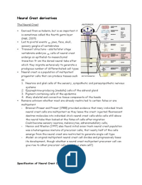
Regionalisation of the Neural crest
Neural crest cells is a transient structure, as its cells undergo E-to-M transition to
disperse throughout body
At different levels of the AP axis – cells centre different tissues and form
different cell types – crest can be divided into 4 but main but overlapping natomical
regions
- Cranial (cephalic) neural crest cells: migrate to produce craniofacial mesenchyme
– differentiates into cartilage, bone, cranial neurons, glia, connective tissues of
face, cells enter pharyngeal arches and pouches to give rise to tooth primordia,
bones of middle ear and jaw
- Cardiac neural crest: Develop into melanocytes, neurons, cartilage, connective
tissue
- Trunk neural crest cells take 2 major pathways: ventrolateral through anterior
half of each somatic sclerotome and differentiate into vertebral cartilage of
each spine, remaining form the dorsal root ganglia containing sensory neurones
(continue more ventrally to form the sympatheitic ganglia, adrenal medulla,
nerve clusters around aorta), 2nd pathway proceeds dorsolaterally – allowing
precursors of melanotcytes to move through to the dermis from the dorsum to
the belly
The Neural Crest
Derived from ectoderm, but is so important it
is sometimes called the fourth germ layer
(Hall, 2009)
Led to pivotal events jaws, face, skull,
sensory ganglia of vertebrates
Transient structure – adults/later stage
vertebrate embryos cells of neural crest
undergo an epithelial-to-mesenchymal
transition fr om the dorsal neural tube after
which they migrate extensively to generate a
prodigious number of differentiated cell types
Neural crest is a population of multipotent
progenitor cells that can produce tissues such
as
1. Neurons and glial cells of the sensory, sympathetic and parasympathetic nervous
systems
2. Epipnephrine-producing (medulla) cells of the adrenal gland
3. Pigment-containing cells of the epidermis
4. Many skeletal and connective tissue components of the heads
Remains unknown whether most are already restricted to certain fates or are
multipotent
- Bronner-Fraser and Fraser (1988) provided evidence that many individual trunk
neural crest cells are multipotent as they leave the crest: injected fluorescent
dextran molecules into individual chick neural crest cells while cells still above
the neural tube then looked at the fates of cells after migration
Could become sensory neurons, melanocytes, adrenomedullary cells,
- Henion and Weston (1997) also found initial avian trunk neural crest population
was a heterogenous mixture of precursor cells, that nearly half of the cells
emerge from the neural crest are restricted to generate single cell type
- Model: an original multipotent neural crest cell divides and progressively loses
its development, though whether a neural crest multipotent precursor cell can
give rise to other precursor cells (ie. a true stem cell?)
Specification of Neural Crest Cells
, Specification is a multi-step process
1. Location of the neural plate border
- Amphibians border appears to be
specified by intermediate
concentrations of BMPs
- Raven and Kloos (1945) showed that
presumptive notochord could induce
both amphibian neural plate and neural
crest tissue (presumably blocking
nearly all BMPs), somite mesoderm and
lateral plate mesoderm could only
induce the neural crest
- In chick embryos, specification occurs
during gastrulation – where borders
between neural and non-neural
ectoderm are still forming (Basch et
al., 2006)
Here, neural plate inductive signals
(especially BMPs and Wnts) secreted from ventral ectoderm and paraxial
mesoderm interact to specify the boundaries
2. In anterior region, timing of BMP and Wnt signal expression is critical for
discriminating between neural plate,
epidermis, placode and neural crest
tissues
- If both BMP and Wnt signalling are
continuous, fate of ectoderm is
epidermal, if BMP antagonists (eg,
Noggin or FGFs) block BMP signalling,
ectoderm becomes neural
- Neural plate induces in these border
cells a set of transcription factors –
neural plate border specifiers –
Distalless-5, Pax3 and Pax7,
collectively prevent border region from
becoming either neural plate or epidermis
- Border specifying transcription factors then induce second set of tfs – neural
crest specifiers in those cells fated to become neural crest (FoxD3, Sox9, Id,
Twist, Snail)
, - When FoxD3, Snail,, Sox3 experimentally expressed in lateral neural tube, these
lateral neuroepithelial cells become neural crest-like, undergo epithelial-
mesenchymal transition, delaminate from then neuroepithelium
- Sox9 and Snail suffiecient to induce transition
- Sox9 is needed for the survival of trunk neural crest cells after delamination (in
the absence, neural crest cells undergo apoptosis as soon as they delaminate)
- FoxD3 plays many roles: needed for expression of the cell surface proteins
needed for migration, critical for specification of the ectodermal neural crest
- Inhibiting FOxD3 gene inhibits neural crest differentiation
- FoxD3 expressed ectopically by electroporating active gene into neural plate
cells, neural plate cells express proteins characteristic of the neural crest
(Cheung et al., 2005)
3. Neural crest specifiers activate transcription of genes that give neural crest
cells migratory properites and some differentiated properties
- Neural crest effectors include some transcription factors (such as MITF in
melanocyte lineage that forms pigment cells), small G proteins (such as Rho
GTPases) that allow cells to change shape and migrate, cell surface receptors
(such receptor tyrosine kinases Ret and Kit) allow neural crest cells to respond
to patterning and inducing proteins in their environment
Regionalisation of the Neural crest
Neural crest cells is a transient structure, as its cells undergo E-to-M transition to
disperse throughout body
At different levels of the AP axis – cells centre different tissues and form
different cell types – crest can be divided into 4 but main but overlapping natomical
regions
- Cranial (cephalic) neural crest cells: migrate to produce craniofacial mesenchyme
– differentiates into cartilage, bone, cranial neurons, glia, connective tissues of
face, cells enter pharyngeal arches and pouches to give rise to tooth primordia,
bones of middle ear and jaw
- Cardiac neural crest: Develop into melanocytes, neurons, cartilage, connective
tissue
- Trunk neural crest cells take 2 major pathways: ventrolateral through anterior
half of each somatic sclerotome and differentiate into vertebral cartilage of
each spine, remaining form the dorsal root ganglia containing sensory neurones
(continue more ventrally to form the sympatheitic ganglia, adrenal medulla,
nerve clusters around aorta), 2nd pathway proceeds dorsolaterally – allowing
precursors of melanotcytes to move through to the dermis from the dorsum to
the belly