Oral Diseases
Classification
● By size: follicle >3mm (cyst), >6mm (cystic change)
● By radiolucency: <15mm (PAG), >15mm (radicular cyst)
● Multilocular: odontogenic cyst (keratocyst, botryoid), odontogenic tumour (ameloblastoma,
myxoma, ameloblastic fibroma, calcifying ep. odontogenic tumour), GC lesion (central GC
granuloma, cherubism, brown tumour of hyperPT), bone cyst (aneurysmal)
● Well-defined, corticated = cysts, benign odontogenic tumour, GC lesions
● Mod well-defined, non-corticated (PO) = chronic infection, surg defect, myeloma
● Poorly defined = acute/spreading infection, malignant tumour
● Effects on adjacent structures: cyst/GC lesion (teeth displaced, no resorption, cortex expansion).
Odontogenic tumour (teeth displaced/resorbed, cortex expansion). Malignancy (teeth
displaced/resorbed, cortex destruction)
Odontogenic Cysts
● “Pathological cavity, fluid/sem-fluid/gaseous contents, not formed by pus accum”
● Ep lining from ep residues of tooth-forming organ
● Developmental (dentigerous, o-keratocyst, DLPD, gingival, glandular-o), Inflammatory (radicular -
AP/LP/residual, paradental)
● Dental lamina → Glands of Serres → o-keratocyst, DLPD, gingival
● Enamel organ → REE → dentigerous
● HERS → Rests of Malassez → radicular
Enlargement of Cysts
● Lysis of epithelial cells, IF cells & IF exudate → high osmotic pressure → fluid flows into cyst
across semi-permeable membrane
● Hydrostatic pressure → cyst enlarges
● Alveolar bone must resorb to allow expansion
● Active agent = prostaglandin
Dentigerous
● Encloses all/part crown of UE tooth, attaches to ACJ
● Eruption cyst = subtype (extra-alveolar, fluctuant bluish mucosal swelling)
● Epid: 2:1 (M:F, Md:Mx), L8 > U3 > U8 > L4/5
● Pres: tooth missing/retained decid
● RG: unilocular, WD RL assoc w/ crown
● Hist: thin ep lining, regular, non-k stratified squamous/cuboidal. Mucous metaplasia common
● Cause:
- fluid b/t REE & enamel?
- outer layers REE prolif → clefts?
- impaction → follicle compression → obstructed venous outflow → increased venous pressure →
increased transudate → increased HS pressure separates follicle & crown
● Tx: XLA tooth, enucleate cyst
Odontogenic Keratocyst (o-keratocyst)
, ● DD: primordial cyst (in place of tooth), keratocystic tumour (mutation/del of tumour suppressor
gene → highly prolif ep lining → aggressive G&R)
● Epid: peak 20/30s, M>F, 75% Md, mainly 8s/4d
● Pres: few symptoms. Enlarge AP. Multiple assoc w. Gorlin-Gotz (BCNS - AD)
● RG: uni/multilocular, WD RL. Can appear dentigerous to L8s
● Hist: thin folded lining, regular stratified squamous ep, thin fibrous capsule. WD basal layer.
Surface layer = parakeratosis (desqaum → cyst lumen)
● Dx: aspirate cyst fluid - desquam ep cells & keratin - lower [solb protein] vs other cysts
● Tx: enucleate/marsupilisation/resection. Hard to remove all lining (10-60% recurrence)
Glandular Odontogenic (glandular-o)
● Epid: 40-50yr
● RG: uni/multilocular, expansive, ANT md
● Effects: may displace, no resorption
Apical Periodontal (Radicular)
● Pres: labial/buccal swelling assoc w/ non-vital tooth. Small; bony/hard. Large; springy, eggshell
cracking, fluctuant, bone erosion
● RG: unilocular, round/oval, WD corticated RL, continuous w/ lamina dura
● APC or PAG: 5-9mm (PAG), 10-14mm (either), APC (>15mm)
● Hist: non-k stratified squamous lining, dense fibrous CT capsule, thin cyst wall, yellow
shimmering cholesterol nodules in fluid (straw-coloured/brown due to haemorrhage)
● Tx: endo tooth/XLA. Doesn’t resolve → enucleate/marsupialise. May remain as residual
,Lateral Periodontal (Radicular)
● Pres: 1cm, round, unilocular
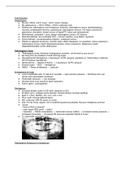
● Botryoid = multilocular version [pic 2]
● Dx: usually assoc with pulp death & non-vital tooth
● Tx: enucleate
Residual
● In apical region of edentulous part of jaw
Paradental
● Cause: IF in PD pocket - usually L8 b/db
● Pres: small, no swelling, history of PC
● RG: WD RL @ neck & coronal ⅓ of root
● Dx: tooth usually vital
● Tx: remove w/ impacted tooth
Inflammatory Collateral Cyst (IF-collateral)
● Epid: kids, 20-40yr
● Pres: B aspect of erupting L6/7/8
● RG: < 4cm, unilocular, WD smooth corticated, assoc tooth tipped
● Tx: XLA assoc tooth +/ enucleate
Calcifying Odontogenic Cyst (calcifying-o)
● Commonly ANT, assoc w/ UE tooth/odontome. Matures & calcifies
, Simple/Traumatic Bone Cyst
● Epid: kids/teens
● Pres: posterior md, no effect on adj structures
● RG: unilocular, irregular (upper border arches up b/t teeth), mod WD, lightly corticated
Aneurysmal Bone Cyst
● Epid: teens
● Pres: painless swelling, rare
● Path: GC
● Tx: curettage
Non-Odontogenic Cysts
● Ep lining (if present) from sources other than tooth-forming organ
● Inclusion: nasopalatine (incisive canal, incisive papilla), globulomaxillary, nasoalveolar, median
palatal
● Congenital: thyroglossal duct, lymphoepithelial, dermoid
● No epithelial lining: bone cyst, salivary gland, stromal cyst
Nasopalatine Incisive Canal Cyst
● Cause: ep residues trapped during palataogenesis
● Epid: 40-60yr
● Pres: asymptomatic/show when infected
● RG: ovoid/heart-shape, behind U1s


