The Reproductive System
Gametogenesis
Gametogenesis is the development of ova and sperm cells, the human sex cells or gametes. The
basic process of gametogenesis for either cell starts with multiplication, in which the diploid (46
chromosomes) germ cells divide many times by mitosis, followed by growth of these daughter cells
and finally cell division by meiosis, producing the cells which form gametes in a process called
maturation.
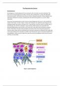
The process of producing sperm cells is known as Spermatogenesis and occurs in the seminiferous
tubules of the testes. As seen in figure 1 below, the male germ cell, called the spermatogonia, first
undergoes growth and mitosis to produce the 1° spermatocyte, and then meiosis to produce the 2°
spermatocyte, this cell then undergoes a second meiosis which produces the spermatid, this
spermatid differentiates into the sperm cell. Sertoli cells in the walls of the seminiferous tubules
secrete a fluid which nourishes the spermatids and protects them from being destroyed by the
immune system; these cells are stimulated by the hormone testosterone released by the Leydig cells
in the spaces between the seminiferous tubules. Once fully formed, sperm cells pass through the 7m
long epididymis, within around 14 days they reach the end of the epididymis where they are stored
until they are ejaculated.
Figure 1: Spermatogenesis
, The male reproductive system is controlled primarily by 4 hormones – luteinizing hormone, follicle-
stimulating hormone, testosterone and inhibin. When a male reaches puberty, the adrenal glands
are able to produce the hormones required to facilitate the production of gonadotropin-releasing
hormone (GnRH) which is controlled by the hypothalamus and acts on the anterior pituitary gland.
GnRH stimulates the anterior pituitary to release follicle-stimulating hormone (FSH) and luteinizing
hormone (LH) into the blood. When FSH is released, it stimulates the Sertoli cells of the seminiferous
tubules to facilitate spermatogenesis. Meanwhile, LH stimulates the Leydig cells which then release
testosterone. Testosterone is responsible for the development of male secondary sexual
characteristics, such as a deeper voice, but also works in a negative feedback loop, as seen in figure 2
below, to inhibit GnRH, FSH and LH. When the sperm count (the concentration of sperm cells in the
semen) is too high, a hormone called inhibin is released from the Sertoli cells into the blood to
decrease the rate at which spermatogenesis occurs. If the sperm count drops too low, around
20million/ml, inhibin is no longer produced and so spermatogenesis is increased (1). The normal
morphology of a sperm cell is an oval head of 4-5um in length 2.5-3.5um in width. There is a ‘cap’
known as the acrosome which allows the cell to penetrate the oocyte, which should cover 40-70% of
the head. The tail should be relatively straight and around 45um long, it is connected to the head by
a midpiece which is less than 1um wide and around 1.5x the length of the head. Sometimes smoking,
alcohol, medications, DNA, chemical exposure and other factors can cause abnormalities in sperm
morphology such as tapered/round/amorphous heads and tails which are too long, too short or
coiled.
Figure 2: Negative Feedback Control of Spermatogenesis (1)
Oogenesis is the process of producing ova, or egg cells. The germ cell of the oocyte is called the
oogonium, this duplicated its DNA to produce the diploid primary oocyte which grows in the ovary
due to FSH. When the follicle grows enough, it releases oestrogen which inhibits FHS to prevent
multiple follicles from developing at once. The primary oocyte then undergoes meiosis to produce a
haploid secondary oocyte and a polar body (unused haploid cell). The secondary oocyte continues to
grow in the ovary where the zona pellucida forms and it becomes known as a Graafian follicle. This
oocyte completes the second stage of meiosis during ovulation, when the follicle ruptures. The
remaining follicle which has released its oocyte forms the corpus luteum which produces
progesterone, a hormone which thickens the endometrium to prepare the uterus for pregnancy.


