College aantekeningen Cognitive Neuropsychology (540033-B-6)
27 views 0 purchase
Module
Cognitive Neuropsychology (540033B6)
Institution
Tilburg University (UVT)
These are lecture notes for the course Cognitive Neuropsychology (-B-6) taken in the year '22/'23 at Tilburg University. The lecture notes contain all lectures except for the guest lecture (lecture 9) which is not mandatory for the exam.
From neurons to electromagnetic field: Excitatory postsynaptic potential (PSP) goes to the dendrites
of the receiving pyramidal neuron which creates an action potential. the dendrites of the pyramidal
neuron become negatively charged and the cell body becomes positively charged which creates a
small dipole and magnetic field
How is EEG measured?:
A stimulus is shown with marker codes, what is measured by the EEG (the raw data) is first filtered
and amplified and then gets digitized to a computer so it is visible. EEG is a high temporal but lower
spatial resolution
The EEG itself measures the voltage difference between 2 electrodes. The result is the active node
minus the reference node which then results in a rhythmic fluctuation in voltage (the wiggles you see
on the screen).
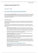
Active, reference, ground nodes: You want to know what the Active – Reference is, to get
there you use (Active – ground) – (Reference – ground). Any noise which is common to both
Active and Reference will be filtered. The purpose of the ground electrode is to reduce noise
According to Luck, the best reference nodes are the mastoids (A1 & A2 picture above), but
others claim that the average of the nodes is the best reference.
EOG (electrooculography): nodes around the eyes which capture left, right, up and down
movements and blinks to filter out that artifact.
Analogue Digital conversion: analogue EEG signals are digitized into voltages at time series
~500hz = 500 measurements per second (per node). The sample rate should be at least 2x
the amount of hertz to withhold it from aliasing.
The AD level sets the resolution of the image or in this case eeg resolution. Think of it that
the true level of voltage in the brain can never be infinitely accurately measured, so there are
intervals at which they can be measured. The higher the resolution, the smaller intervals can
be measured (and thus more accurately)
Major EEG bands: Delta waves (1-3hz) Slow wave sleep, Theta (4-7hz) , Alpha (8-12hz)
internal rather than external focus, Beta (12-30hz) Mentally active and Gamma (30+hz) local
communication
, Michiel de Folter
Event Related Potentials (ERP): EEG changes that are time-locked to sensory, motor or cognitive
events that are used to study correlations.
The different events are marked in the computer and EEG responses in a certain time after the onset
of the stimulus are recorded and marked with the event creating epochs.
The average of all EEG signals of the same event is the resulting Event Related Response.
ERP parameters: Peak to peak; Peak latency; Base to Peak
MEG (Magnetoencephalogram): uses magnetic fields to measure the activity. MEG has a similar
spatio-temporal resolution, but the magnetic field permeates every tissue so there is less smearing of
the signal. MEG is therefore better for the localization of neural sources but is a lot more expensive
Mismatch negativity MMN: is the ERP where you take the deviant response and subtract the
standard response, the result is the MMN.
standard being a beep, and deviant being the response after a higher toned beep, MMN is the
difference in responses which can even be used to measure brain activity of people in a coma.
Lecture 2: Electrophysiological neuroimaging
Peaks != components: The underlying neural sources create the dipole and will therefore be caught
by the nodes on the scalp of the brain. These neural sources however interfere a bit with each other
so the signal that gets caught by the node is a combination of all neural sources.
In short. The peak you see in the ERP is the sum of all underlying brain components. To find the brain
components we encounter the inverse problem…
inverse problem: The inverse problem has to do with neural source estimation. We want to know
which underlying components are responsible for the ERP’s we encounter, but we can not go back
from ERP to neural source, because there are an infinite number of ways to solve the ERP.
Data analysis EEG: Time domain ERP’s: (All about ERP differences in time)
Noise: All electric signals that are not from the brain. (eg. signals from the eyes, heart,
scratching your head etc.)
Inspection and rejection: Looking at the ERP’s and spotting anomalies in the waves and
filtering those segments out.
S/N ratio: Signal to noise ratio, gets lower if there is high artifact rejection, but that also
means a lot of segments will get deleted.
A liberal artifact rejection will make for more trials in the average (because of a low segment
rejection), but there will be more artifacts in the ERP’s and thus a lower signal/noise
, Michiel de Folter
A conservative artifact rejection will yield less trials in the average (because of high segment
rejection), but there will be less artifacts in the ERP’s and thus a higher signal/noise
Topographic interpolation: when 1 or more electrodes are noisy during the entire
experiment, we can estimate that node by taking the average PSP of the surrounding nodes.
Ocular rejection: Delete segments where a blink occurs.
Ocular correction: Is done by using independent component analysis (ICA). ICA unmixes all
the signals from the EEG, takes out the specific (component) signals for eye movements and
then mixes the signals back together.
Filtering: removing higher or lower frequencies from the EEG signal. eg. movement of
electrodes, muscular artifacts, sweating etc. (High-cut/Low-pass filter)
Different filters mean different ERP’s depending on the cut-offs you choose.
Segmentation: cutting the EEG data into segments creating epochs (tiny intervals of time).
segmentation sets the start and ending time of the epoch. eg. 100ms before onset of
stimulus until 700ms after onset of stimulus.
Baseline correction: If you want to compare different nodes with each other or a node with
the average, but one of the 2 has a larger voltage even before stimulus onset the conclusion
“they differ from each other” does not mean a lot when they start from a different place.
They both have to be moved to baseline to make the right comparison.
The baseline correction is basically. taking the average of the node you want to correct, and
subtract the average from the node. This brings it back to baseline. for example the “GO vs
No-GO”-task.
Peak or a-priori: you choose what to do based on previous research and literature
Double dipping: ???
T-test correction: When comparing every time interval of every node for both conditions you
make about (64 electrodes x epoch of 1000ms x sample rate 1000hz = 64 million) T-tests.
High chance of type I errors, False Positives. So use a Bonferoni correction.
Frequency domain: All about Oscillations in a moment
Fourier transformations: Oscillating signals like EEG consist of sine waves of different
frequencies. And the Fourier transformation decomposes the complex sine wave into the
simple sine waves it consists of.
When comparing the frequencies of waves in typical children in comparison with autistic
children, you can see that the relative frequency of certain brain waves are lower than their
typical counterparts.
Frontal-Alpha-Asymmetry: we can compare the left and right hemisphere. Where we look at
the absence of alpha waves, more alpha waves mean less brain activity and vice versa.
The benefits of buying summaries with Stuvia:
Guaranteed quality through customer reviews
Stuvia customers have reviewed more than 700,000 summaries. This how you know that you are buying the best documents.
Quick and easy check-out
You can quickly pay through credit card for the summaries. There is no membership needed.
Focus on what matters
Your fellow students write the study notes themselves, which is why the documents are always reliable and up-to-date. This ensures you quickly get to the core!
Frequently asked questions
What do I get when I buy this document?
You get a PDF, available immediately after your purchase. The purchased document is accessible anytime, anywhere and indefinitely through your profile.
Satisfaction guarantee: how does it work?
Our satisfaction guarantee ensures that you always find a study document that suits you well. You fill out a form, and our customer service team takes care of the rest.
Who am I buying these notes from?
Stuvia is a marketplace, so you are not buying this document from us, but from seller Maggoe. Stuvia facilitates payment to the seller.
Will I be stuck with a subscription?
No, you only buy these notes for £4.71. You're not tied to anything after your purchase.