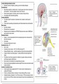
Lesions affecting brainstem function
• Brainstem consists of thalamus, pituitary, pons and medulla oblongata
Pyramidal tract
• Motor fibres originate in cerebral cortex → carries upper motor nerves to spinal cord
and brainstem – here they synapse to lower motor neurones Figure 1 – A diagram
• Fibres transmit signals for voluntary control of muscles of body and face showing the location of the
• No synapses within descending pathway brainstem
Cerebral peduncles
• Two stalks, attach the cerebrum to brainstem and is located in anterior part of
midbrain
• These contain ascending and descending nerve fibres between brain and brainstem
Red nucleus
• Lies within midbrain
• Pale pink due to iron presence
• Receives input from cerebellum of OPPOSITE side and motor cortex of SAME side
and involved in motor control
Brainstem lesions
• Nuclear – rare and associated with other neurological signs due to close proximity Figure 2 – A diagram
with other structures showing the sub
nuclei of the muscles
• Internuclear
supplied by the 3rd
• Infranuclear (below level of nuclei) nerve
Oculomotor nerve – 3rd
• Has 2 nuclei
• Oculomotor nerve nucleus – SR, IR, IO, MR and levator PS
• Erdinger-Westphal nucleus – sphincter pupillae and ciliary body (parasympathetic)
• 2 types of efferent nerve fibres present – somatic (EOM) and visceral (sphincter +
ciliary)
• The efferent nerve fibres originate from midbrain at superior colliculus and leaves
skull through superior orbital fissure
• Each muscle is innervated by its corresponding sub nucleus
• All sub nuclei innervate ipsilateral muscles except SR sub-nucleus and central caudal
nucleus (levators)
Figure 3 – A diagram
Lesions of oculomotor nerve
showing the nerve fibre
• Central caudal nucleus supplies both LPS → lesion results in bilateral ptosis c/s course of the 3rd nerve
unilateral SR limitation
• If there is a bilateral limitation of elevation → lesion affecting SR sub nucleus
• Unilateral limitation of elevation → not SR sub nucleus → SR nerve fascicle involved
as axons from one SR sub nucleus cross and pass through contra and ipsilateral sub
nucleus
,Lesions affecting brainstem function 2
Trochlear nerve – 4th
• Originates in midbrain and exits from posterior midbrain
• Smallest nerve by number of axons but has longest intracranial course
Figure 4 –
• Unable to distinguish between nuclear and fascicular lesions
A diagram
Abducens nerve – 6th showing
• Originates from the paramedian dorsal lower pons in floor of 4th ventricle lateral to the medial longitudinal the
fasciculus nucleus of
the 4th
• Nerve exits at junction of medulla and pons and courses over medial petrous apex towards cavernous
nerve
sinus
Causes of lesions of the 6th cranial nerve
• Brainstem syndrome
• Elevated ICP syndrome
• Petrous apex syndrome
• Cavernous sinus syndrome
• Orbital syndrome Figure 5 –
• Isolated 6th (microvascular) A diagram
Nuclear lesions of 6th nerve showing
• Horizontal gaze palsy where ipsilateral LR and contralateral MR are affected the
nucleus of
• 6th nucleus lies lateral to medial longitudinal fasciculus → some neurones project to MLF and cross over to the 6th
contralateral side and innervate contralateral MR sub nucleus nerve
Fascicular lesions of 6th nerve – ipsilateral LR palsy
Brainstem syndromes
• Caused by lesions such as infarction, haemorrhage, tumour, demyelination, trauma Figure 6 – A
diagram showing
• Causes multiple cranial nerve involvement
the projection of
• Weber’s – midbrain stroke syndrome, 3rd nerve fascicles and cerebral peduncles affected fibres of MLF to 6th
o Ipsilateral 3rd NP and contralateral hemiparesis nerve nuclei,
• Benedikt’s – paramedian midbrain syndrome, 3rd nerve fascicles, red nucleus and cerebral peduncle causing a
horizontal gaze
affected palsy
o Ipsilateral 3rd NP, contralateral hemiparesis, contralateral ataxia with hyperkinesis/tremor
• Foville’s – abducens nucleus, anterior pons and pyramidal tracts affected
o Ipsilateral nuclear 6th NP, ipsilateral horizontal gaze palsy, ipsilateral facial palsy, contralat.
hemiparesis
• Millard-Gubler – base of pons, antero-medially affecting 6th and 7th nerve fascicles and pyramidal tracts Figure 7 –
A diagram
o Ipsilateral 6th nerve palsy, ipsilateral facial nerve palsy, contralateral hemiplegia
showing
Collier’s sign Collier’s
• Unilateral or bilateral lid retraction due to midbrain lesions – characteristic feature of dorsal sign
midbrain/Parinaud’s syndrome
• Also upward gaze palsy, convergence retraction nystagmus, bilateral lid retraction and light-near
dissociation
,Lesions affecting brainstem function 3
Divergence paralysis
• Poorly understood – theories suggest there to be a divergence centre around the 6th Figure 8 – A diagram
showing the 3rd nerve
nerve nucleus and that it is due to a lesion of the cerebellum or Arnold-Chiari
fascicles, red nucleus
malformation and cerebral
• Signs are ET, homonymous diplopia, normal OM but absent negative fusion amplitude peduncles being
• Aetiology is raised ICP, MS, encephalitis, trauma, Miller-Fisher syndrome affected in Benedikt’s
• Differential diagnosis – 6th NP, concomitant ET, convergence spasm
• Treatment – may resolve → observe, occlusion or BO prisms or LR resections as last
resort
Diseases affecting brainstem and OM function
• Parkinson’s – degenerative, insufficient dopamine production – causes are idiopathic,
viral, inherited and drug-induced Figure 9 – A diagram
o Rigidity, tremors are most recognised characteristics – no cure showing the 3rd nerve
o Ocular features – lim. elevation, dep. Affected later, hypometric saccades, CI, fascicles and cerebral
nystagmus, reduced control of phoria, impaired smooth pursuit, blepharospasm, peduncles being
affected in Weber’s
lid lag
• Huntington’s – hereditary, substantia nigra may be involved in brain stem
o Characteristics are loss of mobility, speech and swallowing difficulty
o Ocular features – difficulty initiating saccades, slow saccades, impaired smooth
pursuit
• Wernicke’s encephalopathy – caused by thiamine deficiency, common in alcoholics
and GI disorders Figure 10 – A diagram
o Characteristics are gait ataxia, impaired short term memory – where it is not showing the 6th
nucleus, anterior pons
treated timely, it may progress to Korkasoff’s syndrome which is irreversible
and pyramidal tracts
o Ocular features – abduction weakness, gaze evoked nystagmus, INO, vertical affected in Foville’s
nystagmus, horizontal and vertical gaze palsies, complete ophthalmoplegia
• Whipple’s – Tropheryma whippelii bacteria → weight loss, diarrhoea, GI bleeding, joint
pain etc.
o Treated with antibiotics, bacteria may remain in CSF and fatal if untreated
o Ocular features – reduced vertical saccades, vertical and horizontal gaze palsies
and pendular oscillations
• Arnold-Chiari malformation – anomaly where cerebellar tonsils are displaced Figure 11 – A diagram
downwards towards foramen magnum and may herniate – congenital and acquired showing the 6th and 7th
fascicles, pons and
o Characteristics are head ache, neck pain, tinnitus, facial pain, muscle weakness, pyramidal tracts being
swallowing difficulty, sleep apnoea, impaired coordination, rapid heartbeat, affected in Millard-
dizziness etc. Gubler
o Ocular features – nystagmus especially downbeat, impaired pursuit, impaired
OKN, concomitant ET, divergence paralysis, skew deviation, INO
, Localisation of lesions and life-threatening emergencies 1
• Cerebrum consists of complex neural pathways which process
visual information to coordinate eye movements
• Brainstem is the main cerebral structure containing ocular motor
nuclei and gaze centres
Localisation of function
• Certain areas of the brain are responsible for certain function
• Motor cortex – movement and somatosensory inputs
• Visual cortex – visual information processing
• Broca’s and Wernicke’s area – speech production and
comprehension
Hemispheric lateralisation
• Each hemisphere is specialised to perform certain functions – R
for spatial perception and memory, L for language
Figure 1 – A diagram showing the idea
• Corpus callosum, connects pathways between each hemisphere of hemispheric lateralization – R with
• R side able to compensate for L but L not able to for R → L sided blue and L with red
visual inattention
Function of eye movements
• Eye movements are organised in a hierarchy that descends
down from the most superior cerebral regions to the eyes itself
Figure 2 – A diagram showing the hierarchical
• Pathway is supranuclear centres → brainstem → Infranuclear organization of eye movements, specifically,
pathways → EOM smooth pursuit
Control of eye movements
• Smooth pursuit, saccades, VOR, OKN and vergence
• Pupils and lids
Localisation of OM defects
• Some signs of ocular motility disturbances can localise a lesion
→ indicate affected area Figure 3 – A
• Some signs are non-localising signs and some are false- diagram showing all
localising signs the possible 6th CN
lesions that could
False localising signs – 6th CN
occur and their
• Unilateral or bilateral locations
• Often occurs in context of raised ICP → SOL, idiopathic
intracranial hypertension, cerebral venous thrombosis
• Mechanism → debated, though could be due to long intracranial
course, compression against ridge of petrous temporal bone or
the effect of backwards brainstem displacement