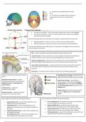
• Frontal sinus is located within the Frontal
Bone
• Skull base has multiple foramina to allow for
bleed vessels and nerves to enter/leave the
skull
• The Anterior Fontanelle – fuses at 18 months post-birth and is known as the BREGMA.
• The Posterior Fontanelle – fuses at 3 months post-birth and is known as the LAMBDA
(looks like Greek letter lambda)
When examining neonates, the fontanelles can be a guide to hydration/intracranial pressure.
• Bulging fontanelle – increase in pressure, may be due to increased hydration
• Depressed fontanelle – decrease in pressure and may be due to reduced hydration
There may be a Metopic suture (10%) which is a continuation of the sagittal suture in the frontal
bone (normally has no suture)
Skin – contains hair follicles and sebaceous glands (may get sebaceous cysts)
Connective tissue – Dense connective tissue which is highly innervated and vascularised so
reduced constriction and can lead to profuse bleeding (has lots of anastomoses)
Aponeurosis (Epicranial) – aponeurosis of the occipitofrontalis muscle, has 2 bellies (frontal (by
orbit) and Occipital (occipital bone)), the pull of this muscle prevents laceration closure
Loose areolar connective tissue – vascularised and contains emissary veins which connect the
veins of the scalp to the diploic veins and intracranial venous sinuses and allows for skin
movement
Periosteum – has superior/inferior tables of compact bone and Diploe (spongy bone)
These are all branches of the External Carotid
Superficial Venous drainage – follows the same
Artery
supply of the blood vessels with the same names
Superficial temporal Artery – supplies
Deep Venous drainage – specifically the
frontal/temporal regions (accompanied by
temporal region, is drained by the Pterygoid
Ophthalmic Artery – Supraorbital and
venous plexus which runs between temporalis
supratrochlear)
and lateral pterygoid muscle and drains to the
Occipital artery – supplies the Maxillary vein
posterior/occipital region
emissary veins which connect the veins of the
Posterior auricular – supplies the superior scalp to the Diploic veins and Dural venous
and posterior auricular (ear) region sinuses
Nerve Supply to the SCALP from the Trigeminal Nerve: Nerve Supply to the SCALP from the Cervical Nerve:
• Supratrochlear nerve – branch of the ophthalmic nerve, • Lesser occipital nerve – derived from the anterior ramus (division)
supplies the anteromedial forehead of C2 and supplies the skin posterior to the ear
• Supraorbital nerve – branch of the ophthalmic nerve, • Greater occipital nerve – derived from the posterior ramus
supplies the scalp between the anterolateral forehead and (division) of C2 and supplies the skin of the occipital region
the vertex. • Great auricular nerve – derived from the anterior rami of C2/3,
• Zygomaticotemporal nerve – branch of the maxillary nerve, supplies the skin posterior to the ear/angle of the mandible
this supplies the temple • Third occipital nerve – derived from the posterior ramus of C3 and
• Auriculotemporal nerve – branch of the mandibular nerve supplies the skin of the inferior occipital region
which supplies skin anterosuperior to the auricle
, The Pterion – the junction of the frontal, parietal, sphenoid and temporal bones
so is a relative weak point of the skull
Trauma to this area leads to an Extra-dural Haematoma due to rapid increase in
intracranial pressure (arterial supply under higher pressure) so strips meninges
and dura mater away from the skull forming lemon shaped bleed
Sub-dural Haematoma – presents as a crescent shaped bleed on an MRI
• Zygomatic (2) – forms the cheek bones of the face and articulates with the
frontal, sphenoid, temporal and maxilla bones.
• Lacrimal (2) – the smallest bones of the face. They form part of the medial wall of
the orbit.
• Nasal (2) – two slender bones that are located at the bridge of the nose.
• Inferior nasal conchae (2) – located within the nasal cavity, these bones increase
the surface area of the nasal cavity, thus increasing the amount of inspired air
that can come into contact with the cavity walls. Superior and Middle conchae
are part of the Ethmoid bone
• Palatine (2) – situated at the rear of oral cavity and forms part of the hard palate.
• Maxilla (2) – comprises part of the upper jaw and hard palate.
• Vomer – forms the posterior aspect of the nasal septum.
• Mandible (jaw) – articulates with the base of the cranium at the
temporomandibular joint (TMJ).
• Supraorbital notch – Allows for the supraorbital notch of the ophthalmic nerve to
pass through
• Infraorbital foramen – Allows for the maxillary nerve to pass through
• Mental foramen – Allows for the Mandibular nerve to pass through
The Temporomandibular Joint (TMJ):
• Contains the articulation between the Mandibular fossa, Articular tubercle
(squamous part of temporal bone) and Mandibular head with articular surfaces
covered by fibrocartilage.
• The articular disk separates articular surfaces so forms 2 synovial joints.
Mandible:
• Alveolar border (superior – teeth) and Base border (Inferior) – Body contains the
Mental Foramen (passage for neurovascular structures (Mental nerve))
• Coronoid process – Attachment for temporalis muscle
• Key for muscle attachment and the Mylohyoid line deperates the floor of mouth
from the neck
• The Inferior Alveolar Nerve (Branch of CNVc) + Artery provides sensation to
teeth/chin and motor function to mylohyoid/A.Digastric
• Lateral ligament – From articular tubule to the mandibular neck. It is a
thickening of the joint capsule, is to prevent posterior dislocation of the
joint.
• Sphenomandibular ligament – originates from the sphenoid spine, and
attaches to the mandible.
• Stylomandibular ligament – a thickening of the fascia of the parotid
gland. Along with the facial muscles, it supports the weight of the jaw.
, Nasal Cavity:
The Nasal cavity is formed by:
• Septal Cartilage (untreated septal haematomas can cause cartilage
necrosis and lead to a saddle nose)
• Perpendicular plate of the ethmoid bone
• Vomer (has Ala posteriorly and nasopalatine groove)
• Nasal crest of maxillary and palatine bones
• Nasolacrimal duct – opens into inferior meatus (inferior to inferior
conchae)
• Semilunar hiatus – contains maxillary ostium (Superior posterior) and
frontonasal duct / anterior ethmoidal air cells
• Ethmoidal bulla – located in the middle meatus
• Posterior Ethmoidal Air Cells – located in the superior meatus
Arterial supply to the Nose and Palate:
• Hard Palate – Supplied by the Greater Palatine Artery/Nerve (Derived
from CNVb) and Nasopalatine nerve (CNVb)
• Soft Palate – Supplied by the Lesser Palatine Artery/Nerve
• Nose – Supplied by the Post./Ant. Ethmoidal artery, and Sphenopalatine
artery, general sensation – CN V, smelling – CN I
All these blood vessels form Kiesselbach`s Plexus (Little`s area), if there is Epistaxis
(Anterior) from this region, more common, the management is as follows:
1. External Compression (First Aid)
2. Cautery
3. Internal Compression (Packing) – rapid rhino`s (Slide on hard palate an
expand – issue is not knowing when to remove)
4. Surgery
Can also provide topical vasoconstriction, reverse coagulopathy and control BP
Posterior epistaxis is generally arising from the sphenopalatine artery
Hutchinson`s Sign – Affects the Nasociliary nerve (CNVa) so in SHINGLES, if
vesicles are present at the tip of the nose – this nerve is affected – this nerve also
supplies the cornea, this may lead to ulceration and loss of sensation
• Incisive fossa – anterior of mouth and transmits the
Nasopalatine nerve (Branch of CNVb) and Greater Palatine
Artery (terminal aspect), however, these run in opposite
directions
• Greater palatine foramen – transmits the greater palatine
nerve
• Lesser palatine foramina – transmits the lesser palatine
nerve
, Skull Base, Foramina and Fossae
• Cribriform Foramina refer to numerous openings in the cribriform plate of
ethmoid bone – allows Olfactory (CNI) nerve axons to communicate with
olfactory bulb
• Only foramina in the Anterior Cranial Fossa
• The Optic Canal allows the Optic nerve (CNII) and ophthalmic artery to pass
through, located in the Middle Cranial Fossa
• Bounded by body of Sphenoid medially and lesser wing of Sphenoid laterally
• The Superior Orbital Fissure allows transmission of oculomotor (CNIII),
Trochlear (CNIV), Ophthalmic (CNVa), and Abducens (CNVI) and
Ophthalmic veins. Located in the Middle Cranial Fossa
• Bordered by lesser wing superiorly and greater wing inferiorly
• Foramen Rotundum allows Maxillary nerve (CNVb) to pass through as
well as an opening to the Pterygopalatine fossa from Middle Cranial
Fossa
• Located at the base of the greater wing of the Sphenoid
• Foramen Ovale allows Mandibular nerve (CNVc) to pass through as well
as an opening into the infratemporal fossa
• Located posterolateral to foramen rotundum in Middle Cranial Fossa
• Foramen Spinosum allows for the passage of the Middle
Meningeal Artery (originates from E.Carotid artery) and Vein
• Located lateral to Foramen Ovale in the Middle Cranial Fossa
• Foramen lacerum is filled by cartilage (dry skull)
• Located at junction of sphenoid, temporal and occipital bones
and in the Middle Cranial Fossa
• Internal Acoustic Meatus connects the Posterior Cranial Fossa
and the inner ear, this allows the Facial nerve (CNVII) and
Vestibulocochlear nerve (CNVIII) along with labyrinthine artery
• Anterior Cranial Fossa has the above borders • Located in the Petrous part of the temporal bone
• Crista Galli is a point of attachment for the Falx Cerebri (fold
of dura matter of the brain) and also separates the Olfactory • Jugular Foramen transmits Glossopharyngeal nerve (CNIX),
bulbs which lie on the Cribriform Plate Vagus nerve (CNX) and Spinal Accessory nerve (CNXI)
• Also has Internal Jugular vein which transverse and sigmoid dural
sinuses drain
• Bound by petrous part of temporal bone anteriorly and occipital
bone posteriorly so is located in Posterior Cranial Fossa
• Hypoglossal Canal transmits the Hypoglossal nerve (CNXII) out
of the Posterior Cranial Fossa next to the Foramen Magnum in
the Occipital bone – has pharyngeal tubercle for attachment of
the Superior Pharyngeal Constrictor
• Foramen Magnum is the largest Cranial Foramen and is in the
Posterior Cranial Fossa, this allows for the passage of the
Medulla and Meninges, Vertebral and A./P. Spinal arteries and
Dural veins
• Middle Cranial Fossa is bound anteriorly by lesser wings and • Accessory nerve (CNXI) ascends through here and joins the
limbus of Sphenoid bone and posteriorly by the Petrous part Cranial division and leaves again through the Jugular Foramen
of temporal bone and dorsum of Sellae of Sphenoid bone
• Carotid Canal is lateral to the Foramen lacerum and • Hiatus of Greater Petrosal Nerve has parasympathetic nerves
Posteromedial to Foramen Ovale and allows for Internal `Hitch-Hiking` with the Facial nerve (CNVII) and it runs anteriorly
Carotid Artery and sympathetic nerves to pass through and collectively forms the Deep Petrosal nerve
• Nerve of Pterygoid canal is formed from the Greater Petrosal
nerve (parasympathetic) and Deep Petrosal nerve (sympathetic)
and opens into the Pterygopalatine fossa


