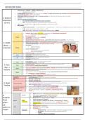
RESPIRATORY EXAM:
• Patient position = URPIGHT > TRIPOD > SUPINE (worst)
o Impending sense of doom?
• SURROUNDINGS: Sputum mug (clear, blood, pus, etc.), inhalers, O2 supply (nasal prongs, non-rebreather), chest physiotherapy devices
(e.g. CF, Bronchiectasis), walking aids, cig packs
• BREATHING WORK: Breathing work @ rest + accessory muscles (esp. intercostal, intraclavicular space, pursed lips)
1. General • Cyanosis (blue lips, blue fingertips)
• Type of cough (dry/productive/bovine),
inspection o Dry = ILD, ACEi, GORD, Post-nasal drip, asthma, sarcoidosis
[at 45o] o Wet = pneumonia, lung abscess, bronchiectasis, APO, TB, lung cancer, PE
• Voice = hoarseness (RLN palsy) – DDx: laryngitis, oeseophageal / lung cancer (+ throat)
• Body Habitus
o Tall thin males (marfan’s) - ++ pneumothorax risk ® elongated blebs + ↑difference in intrapleural pressure b/w apex and base
o T1RF = agitated, tachypnoea, +WOB, confusion ® CPAP
o T2RF = Blue bloaters = flushed skin, bounding pulse, asterixis, drowsy ® BiPAP
• Clubbing = 80% of cases (NOT COPD) ® lung cancer, CF, Bronchiectasis, lung abscess
o Gradual UWL, anaemia, haemoptysis
• Cyanosis (peripheral)
Inspect • Tar staining in-between fingers= SMOKING
• Thinned skin = steroid usage
• Muscle Wasting of small hand muscles ® apical Pancoast’s lung tumour spreading to brachial plexus
invasions of T1 nerve root ® weakening finger abduction/adduction
2. Hands • Wrist tenderness/clubbing = hypertrophic pulmonary osteoarthropathy (HPOA) ® periosteal inflammation
(swelling and tenderness) at the distal wrists [Wrist clubbing]
(distal ® • Fine tremor:
o SABA (B2 agonists)
proximal) Palpate o Steroids / Cushing’s
• Asterixis/Flapping tremor
o (CO2 retention, hepatic encephalopathy)
o Ureamic encephalopathy
• Pamberton’s sign: SVC obstruction
• Tachycardia (asthma, COPD, PE or infection)
Pulse
• Bounding pulse (CO2 retention Type 2 resp. failure)
• Tachypnoea (lung disease, infection, fever, PE, stress)
RR
• Bradypnea (CNS depression)
• Facial plethora—smoker, SVC obstruction [acute SOB]
Face
• Cushingoid appearance – steroid usage
• Conjunctival pallor ® anaemia ® non-respiratory cause of SOB (O2 transport issue)
3. Face Eyes • Horner’s syndrome [miosis, ptosis, anhidrosis]
(Top ® Nose
•nasal polyps (Asthma), engorged turbinates (asthma – allergies), septal deviation (nasal blockage), Sinus
tenderness (palpate maxillary, frontal sinus)
bottom) • peripheral/central cyanosis,
• poor dentition or loose teeth (lung abscess) OR oral candida [UNCONTROLLED ASTHMA]
Mouth
• URTI (red pharynx) + crowded oropharynx
o Check for tonsillitis (URTI), tongue depression required
Warn patient of any discomfort [STERNAL NOTCH = LANDMARK]
Cervical LN • Lymphadenopathy: infection (TB), lung cancer, sarcoidosis
• Raised in cor pulmonale
JVP
• DDx: RVF, TR, PHTN, CCF
• towards side of lung lesion = (atelectasis | fibrosis | pneumonectomy)
4. Neck/ Tracheal deviation
• away from side of lung lesion = (Massive pleural effusion | tension pneumothorax)
• upper mediastinal masses= (Retrosternal goitre)
Trachea o DDx: neck mass – branchial cyst, pharyngeal pouch
• Airflow obstruction è Gross CHEST expansion [possible COPD]
Cricosternal distance &
tracheal tug • Fingers on supraclavicular fossa feel contracted during inspiration (use of
[pull downwards] the accessory muscles – scalenes ® dyspnoea ® impending sign of resp.
Failure)
• Scars (under arms), ® thoracotomy, sternotomy (central), chest drains (MAL)
5. General •
•
Skin changes (erythema/rash), radiotherapy tattoos
Prominent veins (SVC obstruction, caput medusae)
Posterior • Barrel-chest (increased AP diameter = COPD),
• Pectus excavatum (CT disorder)
Chest Inspect Shape • Pectus carinatum (childhood resp. disease – bronchiectasis, CF, asthma)
* Video- /symmetry • Kyphoscoliosis (exaggerated curvature of spine – vertebral TB) - risk of T2RF
(IPPA + assisted • Harrison’s sulcus: Lower rib depression due to severe childhood asthma or
ask thoracoscopic
surgery •
rickets
Mainly upwards = emphysema
patient to (VATS)
• Symmetric, movement for COPD
• Assymetrical ®
cough) Chest wall
movement o Pneumonectomy ® absent BS (initially) – BS present as lung tissue develops
o Rib fracture – Subcutaneous emphysema (crackles under skin)
o fibrosis,
o atelectasis, effusion, pneumothorax
, • Chest expansion (fingers surround
sides of chest BUT do not touch):
o normal ≥5cm (CHECK
posteriorly)
o Hoover’s sign = COPD
(thumbs closer together
during inspiration, and
Palpate further apart in
expiration)
(asymmetry and
reduction of movement) • Feel for RV heave (pulmonary HTN)
• Palpate Ribs/ Costochondral joints
® tender = fractures, prolonged
cough
• Subcutaneous emphysema: diffuse swelling on chest wall or
neck + crackling sensation on palpation (due to air beneath skin
® ? pneumothorax in lung or rib #)
Ask patient to fold their arm or hug pillows
Percuss:
• rotates scapulae anteriorly out of the way [compare L/R]
*Compare both sides
• Percuss bilaterally, NB: liver begins at 5th IC space on right
Pneumothorax Consolidation Pleural Effusion Atelectasis COPD / asthma
[upper lobes] - Pneumonia (collapse)
Tracheal deviation Away None Away Towards None
Chest expansion All reduced ipsilaterally
Vocal Fremitus ¯ ¯
Percussion Hyper-Resonant Dull Stony Dull “blood, Dull Hyper-Resonant
lymph,or fluid” (bullae areas)
Dull (consolidation)
Standard • Patient breaths in/out deeply ® compare both sides for symmetry
Vocal resonance • Repeat auscultation but patients says “99”
1) Breath sound
• Intensity ® reduced diffuse (COPD) or reduced localised (pleural effusion, pneumothorax)
• Quality (vesicular = normal (I:E ratio = 3:1) ® bronchial = periphery (hollow) = consolidated (lobar pneumonia) or
collapsed lung or pleural effusion
2) Added sounds
Auscultate Reduced airway entry (↓BS) Emphysema, pneumothorax, pleural effusion, collapse
(same location as Inspiratory stridor (high pitched • ACUTE: tracheal, laryngeal obstruction (e.g. food, foreign object),
percussion ® compare monophonic) epiglottis, anaphylaxis
both sides) • CHRONIC: laryngeal cancer, tracheal cancer, laryngeal palsy
Expiratory Wheeze (polyphonic vs • COPD [low-pitch polyphonic or monophonic]
*Emergency = ‘silent’ chest monophonic)
= narrowed airway about • Asthma [high-pitch polyphonic
to collapse • Monophonic = Tumour
Coarse crackles (mid-insp, variable) Bronchiectasis, consolidation (Esp. vocal resonance)
Early inspiratory crackles COPD (esp. chronic bronchitis)
Coarse inspiratory crackles Pulmonary oedema, consolidation
Fine-end-inspiratory crackles [Velcro] Pulmonary fibrosis, ILD (upper vs lower lobe)
Pleural rub (grating colicky sound) Pleurisy, pulmonary infraction, pneumonia, pleural malignancy
Whispered petroliquy (transmitted upper Consolidation “sound of words head through chest wall”
airways sounds)
Recognise P2 (loud = pulmonary HT, soft = pulmonary stenosis)
• Loud P2 “ba-boom”
Auscultate Heart • RV heave/thrill
• ↓JVP
• Systolic murmur (TR)
6. Anterior
chest
[Recline
back at
45o]
• Ask to blow hard (FEV1) – check if if > 3seconds
Dynamic • DDx: COPD, asthma or gas trapping
o DD
• Liver = ptosis (hyperinflated chest), hepatomegaly (RVF)
Abdomen • Spllen =splenomegaly (portal HTN secondary to CF or portal vein thrombosis)
+ legs • Peripheral oedema – sacral, tibial, medial malleolus (cor pulmonale)
• Swollen/tender calves (DVT)
Today I performed a respiratory exam on _______ :
• on inspection, any peripheral stigmata of resp. disease? Regular pulse? Normal RR?
Summary • On percussion, symmetrical on front and chest? Any dullness? Any resonance?
• On auscultation, normal vesicular breathing? Added sounds?
• Review observation checks (esp. checking O2 sats and temp. for • FBC, EUC, ABG
To complete the possible infection) • Possible CXR: for possible malignancy or pleural effusion:
examination, I • Send off a sputum sample • CTPA or V/Q sperfusion scan
would like to: • Measure peak flow (if asthmatic) • High res CT scan
• Full Cardiac and Peripheral vascular exam • Lung biopsy
, RESPIRATORY PHYSIOLOGY
Why study the respiratory membrane?
• In emphysema, what effect does the larger size but smaller number of alveoli have on gas exchange?
Less efficient gas exchange
• Why are babies born before 28 weeks at risk from respiratory distress?
Babies have started to produce surfactant to prevent alveolar collapse. No surfactant produced would cause substantial alveolar collapse
and causing respiratory distress.
Ø Give steroids for lung maturity
Ø Surfactant and caffeine to help with respiratory distress
In a non-valvular pneumothorax, why doesn’t the other lung collapse as well?
• Hole is made into chest ® air gets into pleural cavity ®lung collapses due to elasticity and negative pressure lost (Pip =0)(, allowing air to
enter IN and OUT (no valve)
• Remaining lung is not collapsed to keep person alive as there is a midline partition (created by CT from anterior heart to sternum and from
posterior part of pericardium back to spine) to separate the left and right pleura cavities (so air entering in one pleura does not enter to the
other pleura)
• Valvular pneumothorax = flap of tissue created to allow air in but cannot get out. Inspiration allows more air in creating more tension and
pressure to push heart against good lung
• Tension pneumothorax = 14G needle decompression in 2nd IC MCL (1ST LINE) ® 5th IC space AAL
Why doesn’t the lung completely collapse when you breath out (i.e. completely exhaust ERV)?
• Since there is a residual volume (» 1L), which is essential to prevent lung collapse
What’s the deal with foetal haemoglobin?
Foetal haemoglobin holds oxygen very tightly and is able to attract oxygen from
maternal haemoglobin in order to deliver oxygen to the foetus for tissue metabolism
Ø HbF > HbA (causes physiological jaundice in 1st day of life)
Why is carbon monoxide so bad?
• CO has a greater affinity than oxygen to haemoglobin (close to irreversible
binding), hence decreases the number of oxygen binding sites in red blood cells
causing hypoxia (ironically appear bright red) è 100% o2 sats
• Deliver oxygen at very high partial pressure to mitigate this (Dalton’s Law)
• Shifts curve LEFT + DOWN
What the effect of increasing their 2, 3 DPG in anaemia?
• Typically 2,3 DPG disrupts binding of oxygen [increasing its release]
• This is however, useful for anaemia as it allows oxygen to released more easily to
tissues to compensate for the low number of red blood cells.
Why study breathing in and out useful?
Why is someone with peritonitis not going want to take a breath in?
During inspiration, diaphragm moves down ® moves liver down ® hits and moves intestines. If you have inflamed membrane
(PERITONEUM) around intestines, it will hurt when it moves (hence there is only shallow breathing)
Does it matter that post-abdominal surgery patients don’t want to cough?
Despite coughing hurting, coughing is essential to remove any accumulation of mucus or fluid = minimise chance of respiratory infection
(e.g. post-op pneumonia) è aspiration pneumonia
Why do people with breathing difficulties often lean forward and grasp something immobile with both hands?
Mobilies upper limbs to provide extra assistance in inspiration from accessory respiratory muscles (e.g. scalenes). This is done
unconsciously


