UOB: 21005866
Comparative osteology:
Faunal remains portfolio
Introduction
This portfolio includes 2 sections, the first section reflects a laboratory session in which bones from
human and animal specimens were observed and compared. The second section reflects a second
laboratory session in which modifications on bone samples were observed, this section includes a
description of 3 samples observed and the modifications they showed.
Section 1: Reflection of laboratory session (15/03/23)
During this session, I learnt that some animal bones are surprisingly different to humans in shape,
size and function, for example, the metacarpals in cattle (and other nonbipedal mammals) are
significantly thicker than that of humans and are functioned for weight-bearing more than that of
humans (can be seen in figure 8 and 9).
1.1- The femur bones
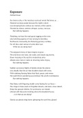
In animal femur bones, there are two faint projections on the inferior portion which were observed
to be significantly more pronounced than in human femurs (can be seen on a cow and human
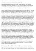
femur in figure 2), in addition, some animals (such as horses) have more projections in other areas
of the bone, which can be seen in figure 1 with a comparison to a human femur. In cows, the
projections split forming a hoof-like shape on the inferior portion of the bone (see figure 2). In
human femur bones, there is a more significant ball projection where the ball and socket joint is
located (can be seen in figures 1 and 2 on the human femur (left bone)), this is not actually fully
present in some animal bones (like in horses) (see figure 1).
Figure 1 – image comparing a Figure 2 – image comparing
horse femur to a human femur cow femur to a human femur
(Adams and Crabtree 2009) (Adams and Crabtree 2009)
, UOB: 21005866
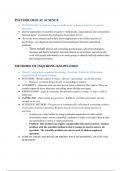
1.2– The vertebrae
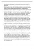
The vertebras of most animals have a significantly longer spinous and transverse process than
humans, this was observed in the laboratory session. Figure 3 shows an image taken from the
laboratory session, where the transverse and spinous processes are missing, this was noted as
being significant as it is a modification (see section 2). It was noticed in the session that the smaller
the animal then the smaller the transverse foramen and vertebral body.
Figure 3 – believed to be a cows Figure 4 – image showing the 7th
vertebrae bone, taken from the thoracic vertebrae of humans (White
laboratory session (Author’s own) and Folkens 2005)
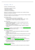
1.3 – The ulna and radius bones
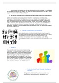
In the lab, I observed and noted that the ulna and radius are both adapted for locomotion in cows
and as a result, these bones take a different shape and thickness to humans (as seen in Figure 5).
Furthermore, the ulna and radius of a cow are fused (can be seen in Figure 5), possibly to add
support for the locomotion of the mammal. A final point is that the projections of the olecranon in
cows are significantly bigger and flatter than in humans (See figure 5). The radius in humans takes a
flat and rounded shape on the superior aspect (seen on the bone in the left of figure 5) but in some
animals, such as cows (seen on the right bone in Figure 5) take a similar shape to a human tibia. The
most interesting to me was the aspects of such bones being able to fuse together. I found that
when looking at the radius of a cow, it was significantly thicker and wider than a human radius and
widens on the inferior aspect of the bone (See figure 5).
Figure 5 – comparison of a human ulna
and radius to a cow’s ulna and radius
(Adams and Crabtree 2009)
Comparative osteology:
Faunal remains portfolio
Introduction
This portfolio includes 2 sections, the first section reflects a laboratory session in which bones from
human and animal specimens were observed and compared. The second section reflects a second
laboratory session in which modifications on bone samples were observed, this section includes a
description of 3 samples observed and the modifications they showed.
Section 1: Reflection of laboratory session (15/03/23)
During this session, I learnt that some animal bones are surprisingly different to humans in shape,
size and function, for example, the metacarpals in cattle (and other nonbipedal mammals) are
significantly thicker than that of humans and are functioned for weight-bearing more than that of
humans (can be seen in figure 8 and 9).
1.1- The femur bones
In animal femur bones, there are two faint projections on the inferior portion which were observed
to be significantly more pronounced than in human femurs (can be seen on a cow and human
femur in figure 2), in addition, some animals (such as horses) have more projections in other areas
of the bone, which can be seen in figure 1 with a comparison to a human femur. In cows, the
projections split forming a hoof-like shape on the inferior portion of the bone (see figure 2). In
human femur bones, there is a more significant ball projection where the ball and socket joint is
located (can be seen in figures 1 and 2 on the human femur (left bone)), this is not actually fully
present in some animal bones (like in horses) (see figure 1).
Figure 1 – image comparing a Figure 2 – image comparing
horse femur to a human femur cow femur to a human femur
(Adams and Crabtree 2009) (Adams and Crabtree 2009)
, UOB: 21005866
1.2– The vertebrae
The vertebras of most animals have a significantly longer spinous and transverse process than
humans, this was observed in the laboratory session. Figure 3 shows an image taken from the
laboratory session, where the transverse and spinous processes are missing, this was noted as
being significant as it is a modification (see section 2). It was noticed in the session that the smaller
the animal then the smaller the transverse foramen and vertebral body.
Figure 3 – believed to be a cows Figure 4 – image showing the 7th
vertebrae bone, taken from the thoracic vertebrae of humans (White
laboratory session (Author’s own) and Folkens 2005)
1.3 – The ulna and radius bones
In the lab, I observed and noted that the ulna and radius are both adapted for locomotion in cows
and as a result, these bones take a different shape and thickness to humans (as seen in Figure 5).
Furthermore, the ulna and radius of a cow are fused (can be seen in Figure 5), possibly to add
support for the locomotion of the mammal. A final point is that the projections of the olecranon in
cows are significantly bigger and flatter than in humans (See figure 5). The radius in humans takes a
flat and rounded shape on the superior aspect (seen on the bone in the left of figure 5) but in some
animals, such as cows (seen on the right bone in Figure 5) take a similar shape to a human tibia. The
most interesting to me was the aspects of such bones being able to fuse together. I found that
when looking at the radius of a cow, it was significantly thicker and wider than a human radius and
widens on the inferior aspect of the bone (See figure 5).
Figure 5 – comparison of a human ulna
and radius to a cow’s ulna and radius
(Adams and Crabtree 2009)