Unit 8: Physiology of Human Body Systems
B: Understand the impact of disorder on the physiology of the lymphatic system and the associated corrective
treatments.
Impact of lymphatic disorders and associated treatments
As part of my college course, I was offered the chance to participate in a one month placement programme which
has been organised in partnership with a national charity who work to engage science students in aspirational work
experience opportunities. On completion of the placement, my learning mentor has asked me to produce a portfolio
of evidence demonstrating the knowledge and understanding I have gained throughout the programme which will
contain a detailed patient case study relating to the normal functioning of the lymphatic system, impairment of
normal function due to lymphatic diseases and the treatments used to correct the effects of lymphatic diseases.
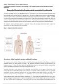
The lymphatic system is the name given to a system of vessels, cells and organs that carries excess fluids to the
bloodstream and filters pathogens from the blood [1].
Figure 1- Diagram of lymphatic system [2]
Structures of the lymphatic system and their functions
The lymphatic system consists of lymph, lymphatic vessels, lymphatic organs (lymphatic nodes, thymus and spleen),
lymphatic nodules and bone marrow [3]. Lymph is the interstitial fluid which is drained into the vessels of the
lymphatic system; it enters the blood system through lymphatic channels and ducts [4]. Lymph maintains fluid
balance and it keeps you healthy by removing bacteria from tissues [4].
Lymphatic vessels are a major component of the lymphatic system and there are two types of these vessels: small
vessels and larger vessels [5]. The smaller vessels are known as lymphatic capillaries and these are made up of a thin
endothelium without a basement membrane [5] The larger lymphatic vessels have muscles in their walls which
helps them gently and slowly pulsate [6]. These vessels also have valves that prevent the backflow of lymph [6].
Lymph nodes are small glands which produce lymphocytes [7]. These nodes are located in the chest, neck, axilla,
abdomen and groin. Lymphocytes are a type of white blood cell which help the body to fight disease and infection
[8]. There are two types of lymphocytes: B cells and T cells [8]. B cells make antibodies which target viruses and
, Unit 8: Physiology of Human Body Systems
B: Understand the impact of disorder on the physiology of the lymphatic system and the associated corrective
treatments.
bacteria [8]. T cells control the body’s immune system response by directly attacking infected cells and tumour cells
[8].
The thymus produces T cells which are distributed throughout the body [6]. It is located in the ribcage just behind
the breastbone [6]. It contains two subcomponents (cortex and medulla) and is composed of epithelial, dendritic,
mesenchymal and endothelial cells [9].
The spleen is the largest organ in the lymphatic system and is located on the left side of the abdominal area
underneath the diaphragm [6]. It contains macrophages, which repair and regenerate damaged tissues, as well as
many cells such as a range of white blood cells, which defend the body from infection [6]. The spleen also destroys
old or damaged red blood cells [6]. It can also help in increasing blood volume quickly if a person loses a lot of blood
[6].
An example of a lymphatic nodule in the lymphatic system is the tonsils, which are located along the inner surface of
the pharynx [10]. They are important in developing immunity to oral pathogens [10]. At the back of the throat are
the pharyngeal tonsil, which can become swollen when responding to an infection [10].
Lymph nodes are small, bean-shaped structures that filter lymph and store white blood cells, which aid in infection
and illness resistance [11]. Lymph nodes are located throughout the body along a network of lymph vessels [11].
These include the axillary nodes (located in the underarm), abdominal nodes (located in the abdomen), inguinal
nodes (located in the groin), popliteal nodes (located behind the knee) and supratrochlear nodes (located in the
medial portion of the forearm).
The axillary lymph nodes are a group of twenty to thirty large lymph nodes located in the deep tissues in and around
the armpit [12]. These nodes are arranged into five groups: pectoral, lateral, subscapular, central and subclavicular
[12]. The pectoral group is made up of four or five large lymph nodes that are found on the superior border of the
pectoralis major muscle [12]. These lymph nodes receive lymph from afferent lymphatic vessels in the breast and
pectoral areas of the chest [12]. The lateral group borders the lateral edge of the pectoral group and contains four to
six lymph nodes clustered around the axillary vein [12]. Lymph from upper limb (arm) lymph vessels enters the
lateral group and travels to the central lymph nodes through efferent lymphatic vessels [12]. The subscapular group
is located in the axilla's posterior region, inferior to the scapula, or shoulder blade [12]. This group consists of six to
seven lymph nodes that filter lymph from lymphatic veins in the back of the neck and upper back [12]. Efferent
lymphatic vessels from this group carry lymph to the central lymph nodes [12]. The central nodes are a group of
three to four lymph nodes embedded in the adipose tissue mass at the base of the axilla [12]. They filter lymph that
has already passed through the pectoral, lateral and subscapular lymph nodes [12]. Lymph from the central nodes
travels through lymphatic vessels to the subclavicular nodes, which are located slightly below the clavicle, or collar
bone [12].
The abdominal nodes are located in front of the lumbar vertebral bodies near the aorta and consist of three groups:
preaortic, paraaortic and retroaortic lymph nodes [13]. Preaortic lymph nodes are found in front of the aorta and
can be divided into coeliac, superior mesenteric and inferior mesenteric nodes [13]. Coeliac nodes drain the gastric,
hepatic and pancreaticosplenic nodes [13]. Both superior and inferior mesenteric nodes drain the mesenteric nodes
[13]. Paraaortic lymph nodes drain kidneys, adrenal glands, ureter, posterior abdominal wall, testis, ovary, uterus
and uterine tubes. Retroaortic lymph nodes drain the peripheral nodes of the paraaortic group found near the
posterior abdominal wall [13]. The inguinal lymph node contains superficial and deep lymph nodes [14]. These
nodes drain the anal canal (below the pectinate line), the skin below the umbilicus, the lower extremity, the
scrotum, vulva, glans penis and the clitoris. [14]


