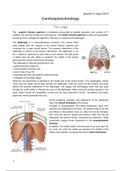
The Lungs
The superior thoracic aperture is completely surrounded by skeletal elements, and consists of TI
vertebra, rib I and the manubrium of the sternum. The inferior thoracic aperture is large and expandable,
formed by bone, cartilage and ligaments. Inferiorly it is closed by the diaphragm.
The diaphragm is a musculotendinous structure. The muscle fibres
arise radially from the margins of the inferior thoracic aperture and
converge into a large central tendon. The posterior attachment of the
diaphragm is inferior to the anterior attachment. The diaphragm is not
flat, it “baloons” superiorly on both sides to form domes. The right dome
is higher than the left. When it contracts, the height of the domes
decreases the volume of the thorax increases.
The diaphragm is attached peripherally to the:
▪ xiphoid process of sternum
▪ costal margin of thoracic wall
▪ ends of ribs XI and XII
▪ ligaments that span the posterior abdominal wall
▪ vertebrae of the lumbar region
Moreover, the pericardium is attached to the middle part of the central tendon. The oesophagus, inferior
vena cava and vagus nerves pass through the diaphragm, while the aorta and the thoracic duct pass
behind the posterior attachment of the diaphragm. The azygos and hemiazygos veins may also pass
through the aortic hiatus or through the crura of the diaphragm. Other structures running laterally to the
aortic hiatus include the sympathetic trunks and the least splanchnic nerves. The greater and lesser
splanchnic nerves penetrate the crura.
During breathing, elevation and depression of the diaphragm
alters the vertical dimension of the thorax.
Changes in anteroposterior and lateral dimensions result from
elevation and depression of the ribs. The anterior ends of the ribs
are inferior to the posterior ends, so, when the ribs are elevated,
they move the sternum upward and forward. When the ribs are
depressed, the sternum moves downward and backward. These
movements change thorax dimensions in the anteroposterior
direction.
In addition, the middle shafts of the ribs tend to be lower than the
two ends. So, when the shafts are elevated, the middles of the
shafts move laterally, increasing the thorax’s lateral dimension.
1
, giovedì 21 marzo 2019
THE PLEURAE
The mediastinum is a thick midline partition of the thoracic
cavity. The two pleural cavities are situated on either side of it
and surround the lungs. Superiorly, they extend above rib I into
the root of the neck, while inferiorly they extend to a level just
above the costal margin. The medial wall of each pleura is the
mediastinum.
Each pleural cavity is lined by a single layer of flat cells,
mesothelium and supporting connective tissue. The pleura is
divided into:
• Parietal pleura, associated with the walls of the pleural cavity
• Visceral pleura, lining the surface of the lungs
The pleural cavity is the tiny space between the two, filled with
a thin layer of serous fluid, which allows the parietal and
visceral pleurae to slide freely.
The parietal pleura is divided into various parts:
▪ Costal part, related to ribs and intercostal spaces.
▪ Diaphragmatic part, covering the diaphragm.
▪ Mediastinal part, covering the mediastinum. In the region of TV to TVII it reflects the mediastinum as a
tubular covering for the structures passing between the lungs. This covering forms the root of the lungs,
which joins the medial surface of the lung to the hilum of lung. In this area the mediastinal pleura si
continuous with the visceral pleura.
▪ Cervical pleura, lining the cervical extension of the pleural cavity. Covering the superior surface of the
cervical pleura is the suprapleural membrane. Made of connective tissue, it is attached laterally to the
medial margin of the first rib and behind to the transverse process of vertebra CVII. Superiorly, it receives
muscle fibres from the scalene group, keeping the membrane taught.
The peripheral reflections of the parietal pleura mark the extent of the pleural cavities. Superiorly, the
pleural cavity projects for 3-4 cm above the first costal cartilage. The
lungs do not completely fill the pleural cavities. This results in
recesses in which two layers of parietal pleura become opposed.
Expansion of the lungs into these spaces occurs only during forced
inspiration. The recesses also provide potential spaces in which fluids
can collect and from which fluids can be aspirated. Anteriorly, a
costomediastinal recess occurs on each side, where costal pleura is
opposed to mediastinal pleura. The largest is on the left side,
overlying the heart. The costodiaphragmatic recesses are the
largest recesses, occurring between the costal pleura and
diaphragmatic pleura.
2
The benefits of buying summaries with Stuvia:
Guaranteed quality through customer reviews
Stuvia customers have reviewed more than 700,000 summaries. This how you know that you are buying the best documents.
Quick and easy check-out
You can quickly pay through credit card for the summaries. There is no membership needed.
Focus on what matters
Your fellow students write the study notes themselves, which is why the documents are always reliable and up-to-date. This ensures you quickly get to the core!
Frequently asked questions
What do I get when I buy this document?
You get a PDF, available immediately after your purchase. The purchased document is accessible anytime, anywhere and indefinitely through your profile.
Satisfaction guarantee: how does it work?
Our satisfaction guarantee ensures that you always find a study document that suits you well. You fill out a form, and our customer service team takes care of the rest.
Who am I buying these notes from?
Stuvia is a marketplace, so you are not buying this document from us, but from seller Greta96. Stuvia facilitates payment to the seller.
Will I be stuck with a subscription?
No, you only buy these notes for £2.35. You're not tied to anything after your purchase.