Anatomy of nasal cavities, pharynx (nasopharynx, oropharynx and laryngopharinx), larynx, caritalges and ligaments of the larynx, intrinsic muscles, infrahyoid and suprahyoid muscles.
Respiratory Tract
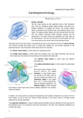
NASAL CAVITIES
The two nasal cavities are the uppermost parts of the respiratory
tract. They are elongated wedge shaped spaces, held open by a
skeletal framework of cartilage and bones. The smaller anterior
region is enclosed by the external nose, and anteriorly opens in the
nares. The larger posterior regions are more central within the skull,
with two posterior apertures called choanae, opening into the
nasopharynx. The nasal cavities are separated from each other by
the nasal septum, from the oral cavity by the hard palate and from
the cranial cavity by the frontal, ethmoid and sphenoid bones.
The lateral wall of the nasal cavity is characterised by three curved shelves of bone, the nasal conchae.
The conchae increase the surface area of contact with respired air, and provide openings for the
paranasal sinuses. They divide each nasal cavity into four air channels.
• The inferior nasal meatus, to which opens the nasolacrimal duct.
• The middle nasal meatus, to which opens the maxillary sinus, the middle ethmoidal cells and the
fronto-nasal duct, that drains the frontal sinus and the anterior ethmoidal cells.
•The superior nasal meatus, to which open the posterior
ethmoidal cells.
•The spheno-ethmoidal recess, to which opens the sphenoidal
sinus.
Each nasal cavity consists of three
general regions. The nasal
vestibule is a small dilated space,
just internal to the naris, lined by
skin and contains hair follicles. The
respiratory region is the largest
part, rich in neuro-vasculature,
lined by respiratory epithelium. The
olfactory region is small, localised
at the apex of each nasal cavity, lined by olfactory epithelium and contains
olfactory receptors.
In addition, the nasal cavities can adjust the temperature and humidity of respired air by action of the rich
blood supply, as well as trap and remove foreign particles in the mucus, which is normally moved
posteriorly by cilia and swallowed.
The respiratory epithelium is a type of ciliated pseudo-stratified columnar epithelium found lining most of
the respiratory tract. It is not present in the larynx and pharynx. The cells in the respiratory epithelium are
of three main types:
1
, domenica 24 marzo 2019
1. Ciliated cells, columnar epithelial cells with specialised ciliary modifications. Contraction of this cilia
pushes away the mucus.
2. Goblet cells, are columnar epithelial cells that secrete mucus, or epithelial lining fluid (ELF), which
helps maintain epithelial moisture and traps foreign particles and pathogens moving through the
airway.
3. Basal cells are small, nearly cuboidal cells thought to have some ability to differentiate into other cells
types found within the epithelium, responding to injury.
Other cells of the respiratory epithelium include brush cells, which are columnar cells with microvilli that
function as chemoreceptors, and small granule cells (Kulchitsky cells), which have neuroendocrine
function. Certain parts of the respiratory tract, such as the oropharynx, are subject to the abrasive
swallowing of food. To prevent the destruction of the respiratory epithelium in these areas, it changes to
stratified squamous epithelium, which is better suited to abrasion.
Innervation of the nasal cavity is provided by three cranial nerves. The olfactory nerve [I], responsible for
olfaction, the trigeminal nerve [V] which carries general sensation, and the parasympathetic fibres of the
facial nerve [VII], innervating the glands. Sympathetic fibres are derived from the T1 spinal cord levels.
Blood supply is provided by the terminal branches of the maxillary and facial arteries, and from the
ethmoidal branches of the ophthalmic artery.
Bones that contribute to the skeletal framework of the nasal cavities include the ethmoid, sphenoid and
frontal bones, the vomer, and the paired nasal, maxillary, palatine and lacrimal bones, as well as the
inferior conchae. The external nose composes is composed partly by bone and partly by cartilage. Bony
parts are the nasal bones, and maxillary and frontal bones. Cartilaginous support is provided by the major
alar and three to four minor alar cartilages, and a single
septal cartilage in the midline.
Important elements of the airways are the paranasal sinuses.
There are four, the ethmoidal cells, the sphenoidal, maxillary
and frontal sinuses.
The nares are held open by the alar cartilages, septal cartilage
and inferior nasal spine, as well as adjacent margins of the
maxillae. The choanae are oval-shaped openings between the
nasal cavities and the nasopharynx. Unlike the nares, they are
rigid openings, completely surrounded by bone. These are the
horizontal plate of the palatine bone, the medial plate of the
pterygoid process and the vomer and the body of the sphenoid bone.
PHARYNX
The pharynx is a musculofascial half-cylinder that links oral and nasal
cavities to the larynx and oesophagus. It is a common pathway for air
and food. The pharynx is attached to the base of the skull and is
continuous below, at the level of CVI, with the top of the oesophagus.
The walls of the pharynx are attached anteriorly to the margins of the
nasal cavities, oral cavities and larynx. The choanae open into the
nasopharynx, the oropharyngeal isthmus opens into the oropharynx
and the laryngeal inlet opens into the laryngopharynx. Moreover, the
pharynx is related to the posterior one-third of the tongue and the
2
The benefits of buying summaries with Stuvia:
Guaranteed quality through customer reviews
Stuvia customers have reviewed more than 700,000 summaries. This how you know that you are buying the best documents.
Quick and easy check-out
You can quickly pay through credit card for the summaries. There is no membership needed.
Focus on what matters
Your fellow students write the study notes themselves, which is why the documents are always reliable and up-to-date. This ensures you quickly get to the core!
Frequently asked questions
What do I get when I buy this document?
You get a PDF, available immediately after your purchase. The purchased document is accessible anytime, anywhere and indefinitely through your profile.
Satisfaction guarantee: how does it work?
Our satisfaction guarantee ensures that you always find a study document that suits you well. You fill out a form, and our customer service team takes care of the rest.
Who am I buying these notes from?
Stuvia is a marketplace, so you are not buying this document from us, but from seller Greta96. Stuvia facilitates payment to the seller.
Will I be stuck with a subscription?
No, you only buy these notes for £2.35. You're not tied to anything after your purchase.