Major Veins
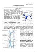
The superior vena cava is a large valveless venous
channel formed by the union of the brachiocephalic
veins. It receives blood from the upper half of the body
(except the heart) and returns it to the right atrium. Its
main tributaries are the azygous vein, and the left and
right brachiocephalic veins.
The azygos vein is a unilateral vessel that ascends in
the thorax to the right side of the vertebral column,
carrying deoxygenated blood from the posterior chest
and abdominal walls. It forms part of the azygos venous
system. The azygos vein is formed by the union of the
ascending lumbar veins and right subcostal veins at
around the level of T12-L2 vertebral level.
If originating at a lumbar level, the azygos vein typically
enters the chest through the aortic hiatus (T12 vertebral level), although may enter by piercing the right
crus. It then ascends in the posterior mediastinum before arching over the right main bronchus posteriorly
at the root of the right lung (at the level of T5-T6) where it joins the SVC. This arch of the azygos vein is
an important anatomical landmark. Tributaries are the hemiazygos vein, a similar structure on the
opposite side of the vertebral column, the accessory hemiazygos vein, the posterior right intercostal
veins, the right superior phrenic vein, right superior intercostal vein, tracheal veins, oesophageal
veins, bronchial veins and pericardial veins. The azygos vein freely anastomoses with the vertebral
venous plexus.
The hemiazygos vein is formed by the
confluence of the left ascending lumbar and
left subcostal veins. It enters the thorax either
through the aortic hiatus or directly through the
diaphragmatic crura. It then courses superiorly
to the left of the midline in the posterior
mediastinum, adjacent to the thoracic vertebrae
until the level of T8 or T9 vertebral bodies,
where it crosses the midline anteriorly to the
vertebral column to drain into the azygos
vein. Tributaries are the left posterior 8th-11th
intercostal veins, left superior phrenic vein,
and occasionally the left renal vein and IVC.
The accessory hemiazygos vein forms part of
the azygos system and along with the
hemiazygos vein, it is partially analogous to the
right-sided azygos vein. It drains the left
superior hemithorax. It is formed by the
1
, sabato 2 marzo 2019
confluence of the middle left posterior intercostal veins. It
descends to the left of midline, adjacent to the thoracic
vertebrae and crosses posteriorly to the aorta at the level of
T7-8 to form a common trunk (interazygos vein) with the
hemiazygos vein to drain into the azygos vein. It normally
anastomoses with the left superior intercostal vein.
Brachiocephalic veins (BCV) drain the head, neck, upper
limbs and part of the thorax and mediastinum. They are formed
by the union of the internal jugular and subclavian veins
posterior to the medial ends of the clavicles.The left
brachiocephalic vein is approximately 6 cm long and runs a
long, oblique course to the right through the superior
mediastinum anterior to the branches of the aortic arch to unite
with the right brachiocephalic vein posterior to the first sterno-
costal joint to form the superior vena cava. Tributaries are the
left vertebral vein, inferior thyroid vein, left internal thoracic
vein, supreme intercostal vein, thymic veins and pericardiophrenic veins.
The right brachiocephalic vein is much shorter, approximately 2.5 cm long and runs a vertical course
anterior to the brachiocephalic trunk. It becomes the SVC as it is joined from the left by the left
brachiocepahlic vein. Tributaries are right vertebral vein, inferior thyroid vein, right internal thoracic
vein and right superior intercostal vein.
The internal jugular vein (IJV) is the major venous return from the brain, upper face and neck. It is
formed by the union of inferior petrosal and sigmoid dural venous sinuses in or just distal to the
jugular foramen, forming the jugular bulb. It descends in the carotid sheath with the internal carotid artery.
The vagus nerve (CN X) lies between the two. It receives tributaries from the face and neck, the
pharyngeal veins, common facial vein, lingual vein, superior thyroid vein and middle thyroid vein. It
continues to descend before descending into the thorax, usually posterior to the space between the two
heads of the sternocleidomastoid muscle, before uniting with the subclavian vein to form the
brachiocephalic vein.
At the level of the lower margin of the orbit, the angular vein
becomes the facial vein. The facial vein runs beneath the
zygomatic muscle, descending along the anterior border and
then on the superficial surface of the masseter. It then crosses
over the body of the mandible, passes obliquely backwards
beneath the platysma and cervical fascia, superficial to the
submandibular gland, the digastric muscle and stylohyoid
muscle. The facial vein pierces
the deep investing fascia of
the neck just below the border
of the mandible where it unites
with the anterior branch of the retro-mandibular vein to form the
common facial vein. The angular vein drains the anterior region of the
scalp. It is formed by the union of the supratrochlear and supraorbital
veins. The supratrochlear vein drains a venous plexus on the anterior
forehead and scalp, while the supraorbital vein drains the anterior part
of the scalp and forehead. The supraorbital vein drains both into the
2
The benefits of buying summaries with Stuvia:
Guaranteed quality through customer reviews
Stuvia customers have reviewed more than 700,000 summaries. This how you know that you are buying the best documents.
Quick and easy check-out
You can quickly pay through credit card for the summaries. There is no membership needed.
Focus on what matters
Your fellow students write the study notes themselves, which is why the documents are always reliable and up-to-date. This ensures you quickly get to the core!
Frequently asked questions
What do I get when I buy this document?
You get a PDF, available immediately after your purchase. The purchased document is accessible anytime, anywhere and indefinitely through your profile.
Satisfaction guarantee: how does it work?
Our satisfaction guarantee ensures that you always find a study document that suits you well. You fill out a form, and our customer service team takes care of the rest.
Who am I buying these notes from?
Stuvia is a marketplace, so you are not buying this document from us, but from seller Greta96. Stuvia facilitates payment to the seller.
Will I be stuck with a subscription?
No, you only buy these notes for £2.35. You're not tied to anything after your purchase.