Summary The Student's Guide to Cognitive Neuroscience (4th ed)
11 views 0 purchase
Module
Neuromarketing
Institution
Erasmus Universiteit Rotterdam (EUR)
A summary of selected chapters from "The Student's Guide to Cognitive Neuroscience", 4th edition, Jamie Ward. This summary contains chapter 2, 3 and 4 as part of the Neuromarketing elective at Rotterdam School of Management, Erasmus University.
Chapter 2 Introducing the brain
Structure and function of the neuron
All neurons have basically the same structure. Consists of
three components:
- A cell body (or soma)
- Dendrites that receive information
- Axon that sends information
Neurons have the same basic structure and function but there are some significant diAerences
between diAerent types of neurons in terms of the spatial arrangements of the dendrites and axon.
The cell body = part of the neuron containing the nucleus and other organelles. The nucleus
contains the genetic code and this is involved in protein synthesis. One of the functions of protein
synthesis is chemical signaling (they can act as neurotransmitters and receptors in neurons).
Neurons receive information from other neurons and they make a “decision” about this information
(by changing their own activity) that can then be passed on to other neurons. From the cell body, a
number of branching structures called dendrites enable communication with other neurons.
Dendrites receive information from other neurons in close proximity. The axon, by contrast, sends
information to other neurons.
Each neuron consists of many dendrites but only a single axon (although the axon may be divided
into several branches called collaterals).
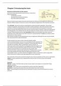
At the terminal of an axon is where chemical signals enable
communication between neurons via a small gap termed a synapse. The
two neurons forming the synapse is called pre-and postsynaptic (before
and after the synapse). When a presynaptic neuron is active, an
electrical current, action potential, is propagated down the length of the
axon. When the action potential reaches the axon terminal, chemicals
(called neurotransmitters) are released into the synaptic cleft.
Neurotransmitters create synaptic potentials. If these passive currents
are strong enough when they reach the beginning of the axon in the
postsynaptic neuron, an action potential is triggered. Passive conduction is short-range while
active conduction enables long-range signaling via action potentials.
The conduction of the action potential along the axon may be speeded up if the axon is myelinated.
Myelin is a fatty substance that is deposited around the axon of some neurons that speeds
conduction.
Keywords
Neuron = a type of cell that makes up the nervous system and supports, among other things,
cognitive function.
Cell body = part of the neuron containing the nucleus and other organelles.
Dendrites = branching structures that carry information from other neurons.
Axon = a branching structure that carries information to other neurons and transmits an action
potential.
1
,Synapse = the small gap between neurons in which neurotransmitters are released, permitting
signaling between neurons.
Neurotransmitters = chemical signals that are released by one neuron and aAect the properties of
other neurons.
How do neurons code information?
The rate of action potential responding, known as the spiking rate, varies among neurons and
relates to the information they carry. The type of information a neuron carries is determined by its
inputs it receives and outputs it sends to other neurons.
For example, the reason neurons in the primary auditory cortex can be considered to carry
information about sound is because they receive input from a pathway originating in the cochlea
and they send information to other neurons involved in more advanced stages of auditory
processing (e.g. speech perception). However, imagine that one were to rewire the brain such that
the primary auditory cortex was to receive inputs from the retinal pathway rather than the auditory
pathway. In this case, the function of the primary “auditory” cortex would have changed (as would
the type of information it carries) even though the region itself was not directly modified (only the
inputs to it were modified). This general point is worth bearing in mind when one considers what
the function of a given region is. The function of a region is determined by its inputs and outputs.
Gray matter, white matter and cerebrospinal fluid
Neurons are organized within the brain to form white matter and gray matter.
- Gray matter consists of neuronal cell bodies.
- White matter consists of axons and support cells (glia).
- Glia support cells of the nervous system involved in tissue repair and in the formation of
myelin (among other functions).
The brain consists of a highly convoluted folded sheet of gray matter, the cerebral cortex, beneath
lies the white matter. In the center of the brain, beneath the bulk of the white matter, lies another
collection of gray matter structures, the subcortex.
White matter tracts may project between
- diAerent cortical regions within the same hemisphere
- diAerent cortical regions in diAerent hemisphere; the most important commissure is the
corpus callosum – a large white matter tract that connects the two hemispheres
- cortical and subcortical structures.
The brain also contains a number of ventricles – hollow chambers of the brain that contain
cerebrospinal fluid (CSF).
2
, There are three di*erent kinds of
white matter tract, depending on
the nature of the regions that are
connected
The brain consists of four ventricles: the
lateral ventricles are found in each
hemisphere, the third ventricle lies centrally
around the subcortical structures and the
fourth lies in the brainstem (hindbrain)
A hierarchical view of the central nervous system
Brain evolution can be thought of as adding additional structures onto older ones, rather than
replacing older structures with newer ones. For example, the main visual pathway in humans
travels from the retina to the occipital lobe, but a number of older visual pathways also exist and
contribute to vision.
Terms of reference and section
Just as with navigation (north, south, east and west) there are conventional directions for navigating
around the brain:
- Anterior and posterior refer to directions toward the front and the back of the brain,
respectively
- Superior/dorsal and inferior/ventral refer to directions toward the top and the bottom,
respectively.
- Lateral and medial refer to directions toward the outer surface and the center of the brain,
respectively.
The terms in the two first bullet points enable navigation in two-dimensions: front-back and top-
bottom.
The cerebral cortex
The cerebral cortex consists of two folded sheets of gray matter organized into two hemispheres
(left and right). The cortex's surface becomes more convoluted over time through evolutionary
progress. The raised surfaces of the cortex are termed gyri (gyrus = singular). The buried
grooves/dips or folds of the cortex are called sulci (sulcus = singular).
3
The benefits of buying summaries with Stuvia:
Guaranteed quality through customer reviews
Stuvia customers have reviewed more than 700,000 summaries. This how you know that you are buying the best documents.
Quick and easy check-out
You can quickly pay through credit card for the summaries. There is no membership needed.
Focus on what matters
Your fellow students write the study notes themselves, which is why the documents are always reliable and up-to-date. This ensures you quickly get to the core!
Frequently asked questions
What do I get when I buy this document?
You get a PDF, available immediately after your purchase. The purchased document is accessible anytime, anywhere and indefinitely through your profile.
Satisfaction guarantee: how does it work?
Our satisfaction guarantee ensures that you always find a study document that suits you well. You fill out a form, and our customer service team takes care of the rest.
Who am I buying these notes from?
Stuvia is a marketplace, so you are not buying this document from us, but from seller weiweihu. Stuvia facilitates payment to the seller.
Will I be stuck with a subscription?
No, you only buy these notes for £2.14. You're not tied to anything after your purchase.