Unit 8A - Physiology of the human body system
The Musculoskeletal System:
The Musculoskeletal system is compromised by many components such as bone, ligaments,
muscles, connective tissues and tendons. The human skeleton is well structured in the body,
it provides structural support to the body, with many other components. The muscles and
skeleton coincide with one another, to maintain posture,
movement and protection to the vital organs.
There are two main parts to the Musculoskeletal System,
the Axial and Appendicular skeleton.
The Axial skeleton provides housing and protection to the
body's vital organs. It is found in the centre of the body,
including the ribs, skull, sternum, spinal vertebrae and
sacrum. It has two main functions, firstly to protect all the
internal organs in the dorsal and ventral cavities, and to
provide a surface for the body's muscles to attach.
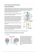
The Axial skeleton is demonstrated in Figure 1 with the
colour blue.
The Appendicular skeleton consists of the body's limb
bones. This includes the clavicle, femur, humerus, pelvis
and many more. This helps the body to be supported in
movement, transfer the weight of the human body, and
acts as a structure to the limbs.
The Appendicular skeleton is demonstrated in Figure 1 with the colour beige.
5 main bones in the Musculoskeletal System and their Functions:
Cranium:
The cranium is eight of the twenty two
bones which compose the entire human
skull. It protects and holds the brain,
along with providing a passage for the
cranial nerves, which are needed for
basic bodily function. The cranial nerves
allow the body to smell, look and eat
food. The cranium is found at the top of
the skull, which joins with other facial
bones.
,During infancy, the cranial bones have significant free spaces in between them, connected
with tissues which feel soft. Once the infant reaches 2-3 months of age, they fuse together.
Mandible:
The mandible is often referred to as the lower
jawbone, it is the strongest and largest facial bone
in the body. The mandible is shaped like a
horse-shoe, and it holds the lower teeth in place.
Maxilla
The maxilla is often referred to as the upper
jawbone. It is responsible for the formation of the
orbit, nose, palate and holding the upper teeth in place.
Additionally, the maxilla plays a vital role in chewing and
speaking.
Vertebral Column
The vertebral column is often known as the spine, it consists of
bony vertebrae which are interconnected by cartilaginous
intervertebral disks. The spinal vertebrae is found from the
base of the skull to the tip of the coccyx. There are 33
vertebrae which make up the spinal column, which are joined
by strong joints and ligaments. It is crucial for the body to bend,
twist and rotate. Additionally, it protects the incredibly fragile spinal cord located in the centre
of the backbone.
There are 5 main parts to the vertebral column,
firstly is the cervical. This part is clinically crucial
as it supplies the arms with nerves, and the
cervical plexus which supplies the diaphragm
with nerves. Additionally, the cervical provides
vital blood flow to the brain. The cervical
consists of the first 7 spinal vertebrae out of the
total 33.
The second part of the vertebral column is the
thoracic. It is composed of 12 spinal vertebrae
and is located in the middle of the spinal
column. It is located in between the cervical and
lumbar vertebrae.
Next is the lumbar vertebrae, consisting of 5
vertebrae. This allows points for appendicular muscles and ligaments to be attached to the
axial skeleton. Additionally, these vertebrae provide stability and mobility at the back of the
spinal column. One of its main functions is protecting the lumbar nerve roots, which branch
, out of the spinal cord, travelling to the buttocks and lower limbs. So, it provides a canal-like
system to protect these vital nerves.
The Sacrum is needed to stabilise the muscles in the legs and abdomen. It is formed by 5
fused bones, which provide the body with leverage during movement and balance, along
with flexibility during childbirth. The sacrum is incredibly strong, it can withstand large
amounts of pressure and motion. It is the main support in allowing the body to sit down.
The Coccyx is made from 4 fused bones, known as the tailbone, and has a triangular shape.
Whilst it is attached to many pelvic floor muscles, it has several redundant and attachment
points. These are incredibly strong and more stable than the coccyx vertebrae. The Coccyx
is the smallest of the vertebral sections, however it is crucial for the body to sit down and
have its weight be supported.
Sternum:
The Sternum is located in the centre of the chest, usually
referred to as the breastbone. It protects the organs from
injury and damage. It is a flat bone which provides
support and protection. It supports the body by acting as a
connection point for the collarbone and rib cage.
Additionally it allows a pivot movement for the skeleton. It
protects the main bodily organs like the earth, lungs and
main arteries in the chest.
Functions of the Musculoskeletal System:
Skeletal muscle attachment:
The attachment will be termed as an origin if the point of attachment is an immobile bone.
Skeletal muscles connect to bones at two ends. Insertion is when attachment is made to an
active bone, in motion during a particular action.
Storing minerals:
In order for the nervous and muscular systems to function properly, the cells within the
skeletal system which release calcium ions into the blood must be successfully maintained.
Red bone marrow stores iron as ferritin molecules and makes haemoglobin from it to carry
oxygen throughout the body. Yellow bone marrow stores lipids as a source of energy to the
body.
Producing blood cells:


