Structural Bioinformatics
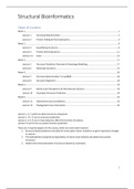
Table of Content
Week 1 ............................................................................................................................................................... 2
Lecture 1 Structural Bioinformatics ................................................................................................... 2
Lecture 2 Protein Folding & Thermodynamics .................................................................................. 4
Week 2 ............................................................................................................................................................... 8
Lecture 3 Quantifying structures ....................................................................................................... 8
Lecture 4 Protein thermodynamics ................................................................................................. 11
Lecture 4.2 Extra ................................................................................................................................. 14
Week 3 ............................................................................................................................................................. 17
Lecture 5 Structure Prediction Overview & Homology Modelling .................................................. 17
Lecture 6 Molecular Dynamics ........................................................................................................ 21
Week 4 ............................................................................................................................................................. 26
Lecture 7 Structure determination: X‑ray/NMR.............................................................................. 26
Lecture 8 Structure Alignment ........................................................................................................ 29
Week 5 ............................................................................................................................................................. 33
Lecture 9 Monte Carlo Simulations for Biomolecular Systems ....................................................... 33
Lecture 10 Secondary Structure Prediction ....................................................................................... 39
Week 6 ............................................................................................................................................................. 42
Lecture 11 Advanced structure prediction ........................................................................................ 42
Lecture 12 Phylogenetic tree construction ....................................................................................... 45
Lecture 1, 3, 7 and 8 are about structure comparison.
Lecture 5, 10, 11 are on structure prediction.
Lecture 2, 4, 6, 9 are on how dynamics affect the function of proteins.
Lecture 12 and 14 are on protein function prediction.
There are 3 required papers for the course, which are main exam material:
1. Structure-based predictions broadly link transcription factor mutations to gene expression changes
in cancers.
2. The hydrophobic temperature dependence of amino acids directly calculated from protein
structures.
3. Atomic-level characterization of structural dynamics of proteins.
1
,Week 1
Lecture 1 Structural Bioinformatics
Read Chapters 0 and 1. Read “Molprobity” paper by Chen et al.
Even though we know full genomes of hundreds of species, our detailed understanding is very limited. In
other words: if we detect a specific mutation in a patient we typically do not know if it is potentially
damaging or harmless.
Dark proteome: part of the proteome for which we don’t know the structure.
Typical structural bioinformatics question: given a sequence, what is its fold? How similar are two
structures? Given a structure, what is its function? How do dynamics affect function?
Core: structure prediction, structure simulation, structure to function and structure comparison.
Protein structure basics
Primary protein structure: side chains.
How can the order of these amino acids determine the shape of the protein?
Special amino acids:
- Cysteine: can create disulphide bridges (covalent bonds); can be a post-translational modification.
- Proline: forms ring with backbone; can have different phi/psi angles.
- Glycine: has no side chain; can have different phi/psi angles.
> Primary structural motifs: alpha helix and beta sheets;
> Secondary structure: helices and sheets. It can be characterised by: planarity of the peptide bond,
favourable hydrogen bonding patterns and steric hindrance.
You can have parallel and anti-parallel beta-sheets.
In what respect are beta strands “less local” than alpha-helices? Can you think of any consequences?
Alpha-helices have 4 amino acid in between them. Two beta-strands can have only a very small loop
between strands, but also a large loop, therefore they are less local.
> Loops tend to be more flexible.
> Hydrogen bonds may be satisfied by: the backbone, a side chain or water.
Secondary structure: planarity of the peptide bond. Favourable hydrogen bonding patterns. Steric
hindrance.
A bond angle VS dihedral angle
Phi-psi angles:
Tertiary structure: how the secondary structure elements connect. You can
characterize it by topology (with SCOP) or architecture (with CATH).
Quaternary structure: interaction with other molecules, and peptide chains
PPI: protein-protein interactions.
PDB: Protein DataBank. It contains X-ray structures, NMR structures, cryo-electron microscopy.
However the PDB contains biases: experimental bias (can we purify, crystallize or stabilize in solution) and
sequence bias.
2
,Knowledge based approaches: data
derived; mined over extended
experimental datasets.
Knowledge based approach ab initio:
from first principles; based on physical
laws.
X-ray crystallography structure
determination >>
Structural ‘genomics’: we are able to
fully sequence genomes. Next is to determine the structure of the entire genome.
It takes most labs nearly 2 years to establish a protein structure with crystallography/NMR.
- Disorder in NMR ensemble; lack of data or protein dynamics?
Transmembrane proteins in PDB; TM proteins are difficult to: express, purify, crystallize and stabilize in
solution.
> Given a pdb file (the coordinates of all the atoms), how would you assign secondary structure?
> Note the difference between secondary structure assignment and prediction. What is the difference?
Measures in PDB file: resolution and R-value.
R-value:
- There is 1 value per structure
- It measures how well the simulated diffraction pattern matches the experimentally-observed
diffraction pattern.
- A totally random set of atoms will give an R-value of about 0.63, whereas a perfect fit would have a
value of 0.
- Typical values are about 0.20.
B-value or Temperature factor:
- One value per atom.
- Measure for the amount of smearing of the electron density.
- Values under 10 create a model of the atom that is very sharp, indicating that the atom is not
moving much and is in the same position in all of the molecules in the crystal. Values greater than
50 or so, indicate that the atom is moving so much that it can barely be seen.
Occupancy:
- One value per atom.
- Some atom groups can be found in multiple confirmations; in this case the atoms are repeated in
the PDB record.
- The occupancy indicates the amount of each conformation that is observed in the crystal.
Are protein structures well represented by the PDB coordinates? What major objections could you have?
Ramachandran plot: only certain combinations of values of phi and psi angles are observed. This is the
situation with main-chain atoms. The Ramachandran plot attempts to bring some order in confirmational
space.
Can we do something similar with side-chain atoms?
3
, Rotamers: highly populated combinations of side-chain dihedral angles.
E.g. Lys ahs 4 angles. The sample amino acid Lysine has four torsion axes within its side chain. The torsion
axes are symbolized as arrows, the dihedral angles are labelled chi1 to chi4.
Side-chains have preferences for discrete parts in space.
The key concept of the lecture: prediction, comparison, simulation and function prediction of proteins;
primary, secondary, tertiary structure of proteins; phi/psi angles; rotamers; h-bonds; bias in PDB and
structural genomics data; structural validation: why and how?
Assignment 1
Part I phi/psi angles from PDB + secondary structure
Part II amino acid propensities
Part III interpretation & propose methods (and read book)
If you have trouble start with part II, 1.1 is probably the most difficult question!
Lecture 2 Protein Folding & Thermodynamics
In the Ramachandran plot only certain combinations of values of phi and psi angles
are observed (only for the backbone).
This is the situation with main-chain atoms. The
Ramachandran plot attempts to bring some order in
conformational space.
Question is whether we can do something similar with the
side-chain atoms? Yes we can, because there also angles
that can rotate
Planes: because the peptide bond does not rotate.
Why are there secondary structures in peptide?
1. planarity of the peptide bond 2. favourable hydrogen bonding patterns 3. steric hindrance.
PDB format:
3 coordinates for the atoms; name of amino acid; position of the amino acid (in this case 1).
The answer of the question below the graph: b
Torsion:
Dihedral:
Dot product: takes two vectors, and transforms
it to a scalar (number)
Cross product: takes two vectors, and transforms
it to a vector of the same dimensions.
n = Normal vector of the plane
4


