Cognitive neuropsychology
Hoorcollege 1
Chapter 1
Cognitive neuropsychology = the study of the relation between structure and function of
the brain and specific cognitive functions. This is done:
- by investigating these cognitive processes in normal healthy people.
- by investigating the breakdown of these processes in brain-damaged individuals.
With imaging methods, you cannot conclude anything about a single person, you can only
draw conclusions on a group level.
Persuasive power of neuro = people are easily convinced that fake neuroimaging
instruments can predict your thoughts.
White matter contains axons. Grey matter contains cell bodies.
Without input (at rest), cell membrane of a neuron has an electrical potential difference
between in- and outside of -70 mV. This is the rest potential. The threshold is at -55 mV.
When it reaches the threshold, the neuron depolarizes. When it comes beneath the -70 mV,
it hyperpolarizes.
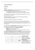
The simplest signal is a sinusoidal oscillation. This signal has a specific frequency. It is 1 Hz in
one second. But in biology, a signal never has only one frequency.
Complex signals can be decomposed into frequency components:
- Amplitude = how much it goes up and down.
- Phase = when it goes up and down.
- Wavelength = the length of one cycle. The longer the wave length, the lower the
activity of the brain. The frequency is 1 : wavelength.
The higher your sampling frequency, the more information you have about the frequency.
For example, if you measure only on second 1 and second 2, the sinus is both times at its
highest and it looks like it didn’t change. However, if you also measure at 1,5 seconds, you
see its at its lowest. The sampling frequency is higher thus you have more information.
The highest frequency is
limited by the sampling
frequency. It is the half of the
sampling frequency (Nyquist
sampling theorem).
The lowest frequency is limited
by how long the signal is
measured. It is 1 divided by the
number of seconds measured.
1
,Filtering = attenuating or excluding certain part of measured frequency spectrum.
- Low-pass, high-pass or band-pass (lower and higher frequencies are excluded).
Smoothing = when the highest or lowest frequencies are not attenuated, but completely left
out of the signal.
Nyquist's sampling theorem = states that a periodic signal must be sampled at more than
twice the highest frequency component of the signal.
Spectogram = the strength of each signal component at each moment in time.
Electrophysiological changes are connected to other kind of changes at different scales:
- At a smaller scale: movement of chemical substances and molecules.
o Depolarization causes an influx of Na+.
o Repolarization causes an outflux of K+.
- At a larger scale: hemodynamics = related to the blood supply which is adjusted to
the current energy needs. If you need more energy, the blood supply is increased.
Active neurons have a high calcium concentration. With two-photon calcium imaging, it is
possible to determine the calcium concentration and therefore a method to measure neural
activity. You can determine the calcium concentration from individual neurons.
Electrophysiological events require energy. The amplitude of potential changes is not
necessarily the best predictor of energy consumption.
Restoring resting potential requires energy. This is the conversion of ATP to ADP + Pi. The
energy consumption of a neuron could correlate with number of action potentials.
Pre- and post-synaptic factors (e.g., neurotransmitter release) also require energy.
Non-invasive methods cannot achieve a single neuron resolution. Even methods with the
highest spatial resolution still average signal from many neurons.
Neurons of similar functional properties are clustered together. The more clustering, the
more the averaged signal from many neurons corresponds to the signal of the individual
neurons. Therefore, the sensitivity of a non-invasive imaging technique depends upon the
amount of clustering present.
An example of clustering are orientation columns. Orientation columns are not present in all
species.
The methods to measure brain structure and activity can be measured in 3 dimensions:
- Temporal resolution = the smallest unit of time that can be differentiated by a
method.
- Spatial resolution = the smallest unit of space which can be resolved.
- Invasiveness = majority of methods are either fully invasive (skull needs to be
penetrated) or not invasive at all.
Most of the methods with a high spatial resolution, are invasive.
2
,Histology = cutting the brain in pieces (e.g. slice of mouse brain) and adding a chemical
substance which colours regions of the brain for visualization of specific structure.
Structural magnetic resonance imaging (MRI) = used for investigation of anatomy in
individuals, anatomical localization of functional findings and relate anatomical structure to
differences between participants.
Hemodynamic methods = you measure changes in blood and tissue oxygenation, blood flow
and blood volume. The temporal resolution of hemodynamic imaging is poorer compared to
electrical imaging due to the slowness of hemodynamic events. The spatial resolution varies
but is smaller than for electrical signals.
Spatial resolution is affected by:
- The distance electrode and the source of the signal. If the electron is next to the
neuron, then the spatial resolution is high. But if the distance is longer, the spatial
resolution will be longer.
- There is an intermediate tissue (the skull).
- Non-invasiveness: the highest frequencies cannot be picked up.
The optimal spatial resolution is possible with patch-clamp recordings, which measures
changes in the potentials with only some little artefacts. The electrode must require a strong
resistance. If the electrode has a smaller resistance, then multi-unit recordings (action
potentials) or local field potentials (LSP) can be measured.
- Multi-unit recordings are often combined with high-pass filtering.
- LSP recording are often combined with a low-pass filter.
Peripheral measures:
- Skin conductance = the index of sympathic arousal intensity in affective or cognitive
processing. Gives an indication of the activation of the SNS.
- Heart activity: gives an indication of the activation of the PNS.
o Heart rate = the time between two beats.
o Heart rate variability = measure the influence of the PNS influence on the
hearts.
o Blood pressure = measure of stress.
- Muscle activity = you can measure with a facial EMG the inferring affective states.
- Eye measure = you can look at saccades (no information processing) or fixations
(information processing) which measure visual attention. Another way is pupil
dilation = gives an indication of the intense emotional arousal towards both pleasant
and unpleasant stimuli and experiences.
Hoorcollege 2
Chapter 9, 10 and 11 (only 11.1.4)
Study the virtual ERP Boot Camp for this lecture chapter 1 till 5.
3
,Electroencephalogram (EEG) = measures the electrical activity of the brain, information
processing. Hans Berger reported the first non-invasive human EEG. He reported that there
were alpha waves when subjects were relaxed and with eyes closed while the alpha waves
disappear with the eyes open and beta waves appear.
Electrodes are attached to the scalp with an EEG. The activity that is measured has to go to
several layers before reaching the neurons.
→ there is no electric current brought into the brain, which is the case with tDCS and tACS.
There are 2 kinds of electrical activity:
- Action potential.
- Post synaptic potential (excitatory or inhibitory).
What is measured by the EEG? It is the post synaptic potential. The action potential is too
short. We measure the dendrites with EEG.
If an excitatory neurotransmitter is released at the apical
dendrites of a pyramidal cell, this will lead to the flow of
positively charged ions into the dendrites, creating
negatively charged area outside of the dendrites. The cell
body is positively charged. Together, the negative and
positive poles create a small electrical dipole. The arrow
goes form negative to positive.
If an inhibitatory neurotransmitter is released at the
apical dendrites, the positive side op de dipole would
point towards the cortical surface.
A dipole can point upwards or downwards. If the neurons
act the same at the same time, all the dipoles add up and
that is what we measure with an EEG. We measure the
aggregated PSPs (the sum of the post synaptic potentials)
at the dendrites of pyramidal cells.
When the direction of the dipole is different for several neurons and you
want to add up all the dipoles, the sum will be zero because the PSPs
cancel each other out.
For every neural source, you always find a positive component on one
side of the head and a negative component on the other side of the
head.
A dipole is an electromagnetic field, so both electrical and magnetical. Electric and magnetic
fields are orientated perpendicular. There is a 90 degrees’ difference. You can measure the
electrical field with an EEG and a magnetic field with an MEG.
4
,It is necessary to relate the task (e.g. the stroop task) to the measurements of the EEG. This
can be done by using marker codes. These codes will then appear in the EEG and give the
exact time at which a stimulus is represented and the time the person gave the answer.
You measure the brain activity with 2 sensors (just like a battery). One electrode is the active
sensor which you place on the part of the brain in which you are interested. The other
electrode is a reference electrode which you place on a relatively neutral part such as the
ear lobes.
→ Important!!
We need a reference electrode as a biological baseline, because organs like the heart
produce much activity which is often even more than signal of the brain activity. The
reference electrode is often placed on the mastoids (M1 and M2 in the 10-20 system).
For each channel you need an active, a reference and a ground electrode.
Ground electrode = reduces noise that comes from the outside of the brain. The
surrounding contains also electrical fields and the ground electrode makes sure this doesn’t
affect the measurement. How does this work?
- First, you measure the voltage between the active and the ground electrode.
→A-G
- Then, you measure the difference between the reference and the ground electrode.
→R–G
- Lastly, we calculate the difference between these two measures.
→ (A – G) – (R – G) = (A – R)
By doing this calculation, you make sure that every noise that A and R have in
common, will be eliminated (because it comes from the outside).
The international 10-20 system = the naming of each electrode based on placement. Each
electrode consists of a letter followed by a number. The letter stands for the lobe on which
the electrode is placed, such as:
- FP = frontal pole.
- F = frontal.
- C = central.
- P = parietal.
- O = occipital.
- T = temporal.
Sometimes an electrode is placed between two regions, for example between the central
and the parietal lobe. Then you use CP, so both letters.
Then, you also have numbers. The odd numbers are located on the left hemisphere and the
right numbers are located on the right hemisphere. The more lateral (towards the ear) the
electrode is located, the higher the numbers. If the electrode is exactly placed on the
midline, then we use the z which stands for zero.
The distance between the electrodes are not absolutely measured but relatively (in
percentage). This is because every head is different in size. The percentages are calculated
by different points on the head:
- Vertex = the highest point of the scalp (pointing upwards).
5
, - Nasion = the nose.
- Inion = the back of the head.
- Preauricular point = the ear (both left and right).
→ you need to know where for example the F4 electrode should be placed based on the 10-
20 system.
Before inserting the electrodes into the cap, there is a gel used to lower the resistance. This
is done because signals have to travel through the skull and the scalp which have a high
resistance. This conductive gel lowers the resistance. The contact point of an electrode is
often made of silver chloride (AgCl).
The ground electrode reduces electrical environmental noise. The reference electrode
provides a biological baseline. The EOG (electrooculography) electrodes monitor the
artifacts which come from the eye movements. The two electrodes which are places above
and beneath your eye measure the vertical movements of the eye and also the blinks, while
the two electrodes near the eye measure the horizontal movements. When you blink, there
is a charge created which is very high and effects the EEG. With the EOG electrodes, we can
correct for this.
An AD box converts analogue signals (microvolts) to digital numbers (0 and 1).
6
,Sampling frequency = the rate of digitization in Hertz.
Sample rate of 500 Hz = 500 x per second means that each 2 ms one sample is taken (a dot).
If the sample rate is high you can recreate the original signal very well. A good sample rate
follows the same line as the analogue signal, as shown below. The dots represent a good
line. If you have a sample rate that is too low, the dots will not represent the correct line,
which is called aliasing.
Nyquist-Shannon sampling theorem = the sample rate should be at least 2x the fastest
frequencies in the signal.
AD level = the amount of information in each sample. If you only have 1 bit, which means
you only have a 0 or a 1. This doesn’t give you enough information. When you have 2 bits,
you have more options, 00, 01, 10 and 11. This gives you more information. The modern EEG
converters use 24 bits.
Event related potentials (ERPs) = EEG changes that are time locked to sensory, motor or
cognitive events. They can track the time course of mental processes.
Epoch = the EEG is simplified in different epochs, which each represent a different stimulus.
For example, if you have a task in which you have to press a button when you see an O, you
have different epochs every time an X or an O is presented. You combine the epoch from all
7
, the X’s and all the O’s. This gives you an average epoch and shows you that the epoch of the
O differs from the epoch from the X.
Each positive (P) and negative (N) peak is followed by a number (e.g. N1, P2, N2, P3, etc.)
representing the distance from the stimulus onset (0 ms).
Steady-state evoked potentials (ssERPs) = the brain response to a repeating stimulus. The
signal to noise ratio of a ssERP is better than that of a normal ERP because it is robust against
latency jitter.s
Grand average = the average across subjects. Everything that is different in each trial, are
cancelled out in the average. Only the things that stay the same can be seen in the grand
average.
The statistical analysis is done using the single-subject waveforms and not the grand
averages.
ERPs provide a continuous measure of activity at each moment in time. It allows us to
measure the brain processes that occur between the stimulus and the response instead of
just measuring the behavioural response.
ERP parameters:
- Peak to peak = from one peak to the next peak.
- Peak latency = when the peak is at its maximum.
- Base to peak = from the peak to the baseline.
- Area = the area underneath the peak, the surface.
The maximal topography = the place on the head where the peak of the ERP is the highest.
But this is not the same as the neural source, which is the brain structure that causes the
activity.
The ERPs are a combination of the neural sources. The waveforms
of the different neural sources add up and give a different scalp
voltages.
MEG = magnetic field permeates (doordringt) biological tissue, fluid and air. Therefore, there
is less distortion and smearing out of the signal. It is better for the localisation of neural
sources, so it has a better spatial resolution than EEG, because there is no resistance of the
scalp, tissue, etc. It therefore consists of many more signals with higher frequencies (such as
gamma) compared to EEG, because it can travel through the skull. It is the less invasive
method of brain imaging. But is costs more than EEG.
Important to remember about MEG is that the magnetic fields are perpendicular.
8


