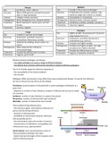
Viruses Bacteria
Size 18-750 nm, most < 200 nm Size Usually 1-20 μm but can be larger
Structure DNA or RNA genome enclosed by protein Structure Prokaryotes (no nucleus); spheres, rods,
shell +/- lipid membrane spirals as single cells, chains, or clusters
Lifestyle ‘obligate cellular parasites’ Lifestyle Extracellular or intracellular
Pathogenesis Direct damage by virus, immune reaction Pathogenesis Bacterial toxins, immune reaction
Treatment Much improved in recent years, but still Treatment Antibiotics; problem antibiotic resistance
limited Prevention Vaccines, hygiene
Prevention Vaccines, hygiene
Parasites
Fungi
Size Highly variable: tiny protozoa (5-10 μm) to
Size Usually 3-5 μm but can be larger large tapeworms (1 m)
Structure Eukaryotes; unicellular (yeast) or Structure Eukaryotes; unicellular (protozoa) or
multicellular-filamentous (molds) multicellular (e.g. worms)
Lifestyle Replicate by budding, fission and/or via Lifestyle Complex and highly variable
spores
Pathogenesis Complex and highly variable; very high
Pathogenesis Often opportunistic infections; prevalence (2.5 billion infected)
immunopathology
Treatment Possible but difficult due to toxicity
Treatment Possible but difficult due to toxicity
Prevention Hygiene, prophylaxis (malaria)
Prevention Hygiene, good health
Relation between pathogen and disease
- one single pathogen can cause a range of different diseases
- one disease (e.g. common cold) can be caused by a range of different pathogens
The % of clinically apparent infections depends on:
- the susceptibility of the host (condition)
- the microbe
Pathogens differ enormously in how often they cause symptomatic disease → overall, the infections
we see as clinical are only the tip of the iceberg
Prevalence: (total) number of infected (with a certain pathogen) individuals at a
given time
- depends on number of new infections, duration of disease and loss due to death
(or recovery)
Incidence: number of new infections in a given time period
Recurrence: number of infected which get reinfected
Mortality: number of infected lost due to death
Factors determining infectiousness
- the infectious agent: time between infection of a
person and becoming infectious
- duration of infectiousness
- probability of transmission between infectious
and susceptible person
- the environment: type and number of contacts
- characteristics of individuals: susceptibility and
infectiousness (e.g. superspreader)
Serial interval: time interval between onset of
clinical symptoms between two cases
- determined by the length of the incubation time
and the infectious period
,- infection control if the serial interval is shorter than the incubation time: it becomes more difficult,
because people are spreading the infection before they know that they are sick
Duration time: time it takes for number of cases to double → exponential growth
- the duration time can vary a lot
- depends on the incubation time of the infection or on the serial interval
Basic reproduction number R0: average number of infected individuals produced by one infected
case (number can vary greatly)
- it is important that the R0 of an infectious disease is reduced to
below 1, because then one person does infect less than one
other person and the infection will die out
Major features of pro- and eukaryotes
1. Prokaryotes
- have a very simple make-up
- do not have a nucleus
- unicellular microorganisms
- have a cell wall
- have little ribosomes in the cytoplasm
- have DNA in the middle of the cell
- also loose plasmids might be present there
2. Eukaryotes
- have cell organelles and have a nucleus
- mitochondria and chloroplasts are coming from bacteria that were
engulfed by some kind of prokaryotic cell that took up these bacteria
or archaea and made them into mitochondria (and chloroplasts)
,- mitochondria have their own ribosomal RNA as well → prokaryotic cell within an eukaryotic cell
- our endoplasmic reticulum (membrane system) contains the ribosomes → especially in the rough
endoplasmic reticulum
Gram stain morphology of bacteria
- gram positive cell wall: made up out of one membrane with on top a whole thick layer of
peptidoglycan cell wall mixed with teichoic acid and lipoteichoic acid → this particular peptidoglycan
can be stained by crystal violet gram staining → stains all bacteria purple, and is fixed by iodine and
then if you decolorize by alcohol or acetone then either the color gets lost (in Gram negative
bacteria) or the color remains (then you have a Gram positive bacteria; color gets stuck in the
peptidoglycan layer)
- safranin red or fuchsine stain the bacteria red or reddish or pinkish
- Gram negative don’t retain the Gram (crystal violet) stain, because they don’t have this
peptidoglycan layer on the surface of the bacteria
- Gram negative have a double lipid layer → 2 membranes: an inner (cytoplasmic) membrane and an
outer membrane; and in the middle there is the perisplasmic space and in there, there is a very thin
peptidoglycan layer and extra proteins
- on top of the Gram negative cell wall there are lipopolysaccharides (LPS), attached to the lipids in
the cell wall
- LPS, if it is taken up by the human body or comes into
bloodstream, is very toxic for us → immediately an immune
response to the LPS
- also look at the form of the bacteria: coccus (round shape),
bacillus (rod shape), coccobacillus (between round and rod
shape), fusiform bacillus, vibrio, spirillum, spirochete
Peptidoglycan network: it is a polymer of N-acetyl-
glucosamine and N-acetyl-muramic acid that are bound in
long chains
- layers of these long chains are bound by pentapeptides (5
amino acid) chain → makes a thick layer
- cleave and kill the bacteria by lysosome treatment (which is also a defence of the human body)
- build-up of peptidoglycan is hampered by antibiotics (e.g. penicillin)
Sporulation: way to produce a bacterial cell with all the genetic
information inside that can survive very harsh conditions
(mostly drought (drying out), or high temperatures)
- not for every bacteria (Gram-negative never sporulate) →
special groups of bacteria can do it (Gram-positives)
- done by engulfment of a cell with an extra cell membrane →
spore has an inner and outer cell membrane
- dipicolinic acid makes the spore very resistant against stress
Spread of antibiotic resistance: bacteria can share their DNA:
1. Transformation
2. Transduction via bacteriophages
3. Conjugation
4. Transposition
, Bacterial metabolism
1. Aerobic or facultative bacteria
- fructose to fructose-phosphate in glycolysis costs 2 ATP
- electrons from the glycolysis go via NADH to another place
- pyruvate gets broken down via citric acid cycle (TCA/Krebs cycle)
- first pyruvate gets decarboxylated, NADH comes free
- acetyl CoA is broken down stepwise → more NADH is made,
some FADH2 (electron carrier) and 2 GTP
- electron carriers go to the electron transport chain to be coupled
finally to oxygen by generating a proton motive force → forms
new ATP via ATP synthase
- one molecule of glucose → 38 ATPs
2. Anaerobic bacteria
- no electron transport chain and usually no citric acid cycle
- bacteria do fermentation: use the glycolysis to splice glucose into
pyruvate (gains 2 ATP), but then they use the pyruvate again to
couple the electrons from NADH back to the reaction products →
pyruvate with the use of NADH is made into lactate or acetate or
into other products
- process gains possibly only 2 ATPs and probably only a few more ATP via the proton motive force
that is also generated in these bacteria
Pathogens: bacteria that cause disease when entering the body
Commensals provide colonization resistance: there are so many bacteria that every corner of our
body is sort of covered by bacteria, so that every pathogen that tries to get in will have to compete
with the bacteria present there
Pathobionts: commensal bacteria that cause disease when giving the opportunity (become from
symbionts and turn into pathogens)
Human barriers
1. Skin
- dry and acidic
- keratin (tough): difficult to degrade (hold
the bacteria outside)
- erosion: shedding of bacteria
- toxic fatty acids: kill bacteria
- microbiota: competition (colonization
resistance)
- antimicrobial peptides (defensins)
2. Mucosa:
- lysozyme: degrades peptidoglycan
- lactoferrin: bind iron → Fe-limitation
- secreted IgA: covalently links all bacteria to themselves and in this way they cannot invade you
- antimicrobial peptides (defensins)
3. Epithelium, gastric acid, gal, digestive enzymes, competition microbiota
4. Innate, antigen-non-specific, immune response
5. Adaptive, antigen-specific, immune response


