100% tevredenheidsgarantie Direct beschikbaar na betaling Zowel online als in PDF Je zit nergens aan vast
logo-home
Summary histology €8,49
In winkelwagen

Samenvatting

Summary histology

2 beoordelingen
 30 keer bekeken  1 keer verkocht

I passed this course with a 7.5

Voorbeeld 4 van de 44  pagina's

  • Nee
  • Chpt 4, page 161 to 191 (4-1 to 4-10) chpt 6, page 229 to 242 (6-3 to 6-6) chpt 10, page 338 to 357
  • 27 december 2021
  • 44
  • 2021/2022
  • Samenvatting
book image

Titel boek:

Auteur(s):

  • Uitgave:
  • ISBN:
  • Druk:
Alle documenten voor dit vak (7)

2  beoordelingen

review-writer-avatar

Door: edaselinkaya • 10 maanden geleden

review-writer-avatar

Door: sapnasafi7 • 1 jaar geleden

avatar-seller
lvd26
Summary Histology
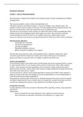
A tissue is a collection of specialized cells and cell products that carry out a limited number
of functions. The study of tissues is called histology. There are four basic tissues:
- Epithelial tissue covers exposed surfaces, lines internal passageways and chambers,
and forms glands.
- Connective tissue fills internal spaces, provides structural support for other tissues,
transports materials within the body, and stores energy.
- Muscle tissue is specialized for contraction and includes the skeletal muscles of the
body, the muscle of the heart, and the muscular walls of hollow organs.
- Nervous tissue carries information from one part of the body to another in the form
of electrical impulses.

Embryonic origin: cells types that are part
of the same basic tissue type can look
much different. What these cells share is
the same embryonic origin.


Epithelial tissue
Epithelial tissue includes epithelia and glands. Epithelia are layers of cells that cover external
or line internal surfaces. Glands are structures that produce fluid secretions. They are either
attracted to or derived from epithelia.
An epithelium is classified based on its embryonic origin (ecto-, endo-, mesoderm),
morphology (the shape of the cells), and cell organization (number of cell layers, strata).

Functions of epithelial tissue:
- Provide physical protection. Epithelia protects exposed and internal surfaces from
abrasion, dehydration, and destruction by chemical or biological agents.
- Control permeability. Any substance that enters or leaves the body must cross an
epithelium. The permeability can differ from cell to cell. The epithelial barrier can
also be regulated and modified in response to stimuli e.g., hormones and physical
stress.
- Provide sensation. Most epithelia are extremely sensitive to stimulation, because
they have a large sensory nerve supply. These sensory nerves provide information
about the external and internal environments. A neuroepithelium is an epithelium
that is specialized to perform a particular sensory function
- Produce specialized secretions. Epithelial cells that produce secretion are gland cells.
In a glandular epithelium, most of all of the epithelium cells produce secretion.
Secretion can provide physical protection or temperature regulation when it is
discharged to the surface of epithelium, but they can also act as chemical messengers
(release in blood or fluid).

,Characteristics of epithelial tissue:
- Polarity refers to the presence of structural and functional differences between the
exposed and attached surfaces. An epithelium consisting of a single layer of cells has
an exposed apical surface and an attached basal surface. In some epithelia, such as
the lining near kidney tubules, mitochondria are concentrated near the base of the
cell, where energy is in high demand for the cell’s transport activities.
- Cellularity. Epithelia are made almost entirely of cells bound closely together by
interconnections known as cell junctions.
- Attachment. The base of an epithelium is bound to a tin noncellular basement
membrane. The basement membrane adheres to the basal surface and to the
underlying tissues to establish the cell’s border and resist stretching.
- Avascularity. Epithelia are avascular, which means that they lack blood vessels.
Epithelia cells get nutrients by diffusion or absorption across either the exposed or
attached surface.
- Regeneration. Epithelial cells that are damaged or lost at the exposed surface are
continuously replaced through stem cell divisions in the epithelium. Regeneration is a
characteristic of other tissues as well, but the rates in epithelia are much higher.

For the epithelium to function as a whole to perform the function just listed, individual cells
may be specialized for:
1. The movement of fluids over the epithelial surface, providing protection and
lubrication.
2. The movement of fluids threw the epithelium, to control permeability.
3. The production of secretions that provide physical protection or act as chemical
messengers.

The specialized epithelial cell is often divided into two functional regions, which means the
cell has a strong polarity. One is the apical surface, where the cell is exposed to an internal
or external environment. The other consists of
the basolateral surfaces, which include both
the base (basal surface), where the cell
attaches to underlying epithelial cells or
deeper tissues, and the sides (lateral surfaces),
where the cell contacts their neighbors.
Microvilli (short cytoskeleton of actin,
microfilaments) are especially abundant on
epithelial surfaces where absorption and
secretion take place, such as along proportions
of the digestive system and kidneys.
A typical ciliated cell contains 250 cilia (long
cytoskeleton of microtubule, tubulin) that
beat in a coordinated manner. It moves
substances over the epithelial surface.

,Maintaining the integrity of epithelia
To be effective as a barrier, an epithelium must form a complete cover or lining. Three
factors help maintain the physical integrity of epithelia:
1. Intercellular connections
2. Attachment to the basement membrane.
3. Epithelia maintenance and repair.

1. Intercellular connections
Large areas of opposing plasma membranes are interconnected by
transmembrane proteins called cell adhesion molecules (CAMs).
Which bind to each other and extracellular materials. Cell junctions
are specialized areas of the plasma membrane that attach a cell to
another cell or to extracellular materials. The three most common
cell junctions are: gap junctions; tight junctions; and desmosomes.

Gap junctions
Some epithelial cells require rapid intercellular communication. At
a gap junction, two cells are held together by two embedded
interlocking transmembrane proteins called connexons. Each
connexon is composed out of six connexin proteins that form a
cylinder with a central pore. Two aligned connexons form a
narrow passageway that lets small molecules and ions pass from
cell to cell. The movement of ions helps coordinate functions as
the beating of cilia.

Tight junctions
Tight junctions encircle the apical regions of epithelial cells. The lipid
portions of the two plasma membranes are tightly bound together by
interlocking membrane proteins. Tight junctions prevent the diffusion of
fluids and solutes between the cells. A continuous adhesion belt lies deep
to the tight junction. This belt is tied to the microfilaments of the terminal
web and is made by transmembrane-linker proteins: cadherins. Tight
junctions effectively isolate the contents of the lumen from the basolateral
surfaces of the cell.

Desmosomes
Desmosomes are very strong and can resist stretching and twisting. Within each cell, a
complex known as a dense area Is connected to the cytoskeleton. This connection to the
cytoskeleton gives the desmosome- and the epithelium- its strength. There are two types of
desmosomes:
- Spot desmosomes are small discs connected to bands of intermediate filaments. The
intermediate filaments stabilize the shape of the cell.
- Hemidesmosomes resemble half of a spot desmosome. A hemidesmosome attaches
a cell to extracellular filament is the
basement membrane. This helps stabilizing
the position of the epithelial cell and
anchors it to underlying tissue.

, 2. Attachment to the basement membrane
Epithelial cells must be attached to other cells, or they will die. The inner surface of each
epithelium is attached to a two-part basement membrane composed of basal lamina and a
reticula lamina. The layer closer to the epithelium, the basal lamina, is an amorphous, ill-
organized layer thought to function as a selective filter. It is secreted by the adjacent layer of
epithelial cells. The basal lamina restricts the movement of proteins and other large
molecules from the underlying connective tissue into the epithelium. The reticular lamina
gives the basement membrane its strength. The reticular lamina also acts as a filter that
determines what substances can diffuse between adjacent tissues and the epithelium.

3. Epithelial maintenance and repair
Epithelial cells are exposed to disruptive enzymes, toxic chemicals, pathogenic bacteria, and
physical distortion. The only way the epithelium can maintain its structure over time is by
continual division of stem cells. Most epithelial stem cells are located near the basement
membrane, in a relatively protected location.


Classification of Epithelia
There are many different specialized cells of
epithelia. You can easily sort these in categories
based on
(1) the cell shape, and
(2) the number of cell layers between the
basement membrane and the exposed surface of
the epithelium.

If only one layer of cells covers the basement
membrane, that layer is simple epithelium. All the
cells have the same polarity, so the distance from
the nucleus to the basement membrane does not
change from one cell to the next. Because they
are so thin, they are fragile. A single layer of cells
cannot provide much mechanical protection, so
simple epithelia are located only in protected
areas inside the body. They line internal
compartments and passageways. Simple epithelia
are also characteristics of regions in which
secretion and absorption occurs, then thinness is
an advantage. It reduces the time required for
materials to cross the epithelial barrier.

On the next page we will discuss the three basic shapes of epithelium cells.

Voordelen van het kopen van samenvattingen bij Stuvia op een rij:

Verzekerd van kwaliteit door reviews

Verzekerd van kwaliteit door reviews

Stuvia-klanten hebben meer dan 700.000 samenvattingen beoordeeld. Zo weet je zeker dat je de beste documenten koopt!

Snel en makkelijk kopen

Snel en makkelijk kopen

Je betaalt supersnel en eenmalig met iDeal, creditcard of Stuvia-tegoed voor de samenvatting. Zonder lidmaatschap.

Focus op de essentie

Focus op de essentie

Samenvattingen worden geschreven voor en door anderen. Daarom zijn de samenvattingen altijd betrouwbaar en actueel. Zo kom je snel tot de kern!

Veelgestelde vragen

Wat krijg ik als ik dit document koop?

Je krijgt een PDF, die direct beschikbaar is na je aankoop. Het gekochte document is altijd, overal en oneindig toegankelijk via je profiel.

Tevredenheidsgarantie: hoe werkt dat?

Onze tevredenheidsgarantie zorgt ervoor dat je altijd een studiedocument vindt dat goed bij je past. Je vult een formulier in en onze klantenservice regelt de rest.

Van wie koop ik deze samenvatting?

Stuvia is een marktplaats, je koop dit document dus niet van ons, maar van verkoper lvd26. Stuvia faciliteert de betaling aan de verkoper.

Zit ik meteen vast aan een abonnement?

Nee, je koopt alleen deze samenvatting voor €8,49. Je zit daarna nergens aan vast.

Is Stuvia te vertrouwen?

4,6 sterren op Google & Trustpilot (+1000 reviews)

Afgelopen 30 dagen zijn er 52510 samenvattingen verkocht

Opgericht in 2010, al 14 jaar dé plek om samenvattingen te kopen

Start met verkopen
€8,49  1x  verkocht
  • (2)
In winkelwagen
Toegevoegd