BBS1004 Brain, Behavior, and Movement - 01/04/22
Case 1 The cerebral cortex
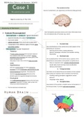
Has Gyri (coils/twists), sulci (grooves), and fissures (deep grooves).
The central nervous system
Macro anatomy of the CNS
The CNS consists of the brain and the spinal cord.
Anatomy of the brain
Each hemisphere processes sensory and motor information from
✽ Forebrain (Prosencephalon)
the contralateral side of the body (e.g. vision)
- Telencephalon or cerebrum (grote hersenen)
→ expands laterally into Lateral hemispheres),.
→ Four brain lobes
→ Lateral ventricles (1st & 2nd) are located here
→ Comprised of two different types of tissue: gray 1. Frontal lobe
and white matter. Lies rostral/anterior of the central sulcus and superior of the
- Diencephalon (tussen hersenen) lateral fissure. Functions:
- Planning future actions, making decisions
○ Thalamus
- Memory, language, motivation, and problem solving
A limbic system structure that connects areas of - Personality
the cerebral cortex that are involved in sensory 2. Parietal lobe
perception and movement with other parts of the
brain and spinal cord. Lies caudal/posterior of the central sulcus, superior of the
Also plays a role in the control of sleep and wake lateral fissure, and anterior of the parieto-occipital sulcus.
cycles. - Plays an important role in somatic sensation/senses
○ Hypothalamus (smell, taste, touch) and eye-hand coordination.
(Sensory information)
Acts as a control center for many autonomic
functions including respiration, blood pressure, and
3. Temporal lobe
body temperature regulation. This endocrine Lies inferior to the lateral fissure and anterior of the occipital
structure secretes hormones that act on the lobe. It plays an important role in emotions and memory.
pituitary gland to regulate biological processes. - Smelling, tasting, perception, aggression, sexual
behavior.
○ Pineal gland
- Language center, speech
This small endocrine gland produces the hormone
melatonin (regulation of sleep-wake cycles and 4. Occipital lobe
influences sexual development)
Lies posterior of the parieto-occipital sulcus and posterior of
the temporal lobe. It controls sight and recognition.
→ The 3rd ventricle is located here
,✽ Midbrain (Mesencephalon)
- Regulates movement and aids in the processing of
auditory and visual information.
○ Tectum
- The cerebral aqueduct is located here. (connects 4th
ventricle to the 3rd ventricle) The dorsal portion of the midbrain that is composed of
the superior and inferior colliculi
(rounded bulges that are involved in visual and
auditory reflexes (before this information goes to
the occipital and temporal lobes).)
○ Cerebral peduncle
The anterior portion of the midbrain consisting of large
bundles of nerve fiber tracts that connect the forebrain to
the hindbrain. Structures of the cerebral peduncle
include:
- The tegmentum
Involved in movement coordination and
alertness
- Cerebral curs (crus cerebri)
Contains the motor tracts, traveling from the
cerebral cortex to the pons and spine.
○ Substantia nigra
✽ Hindbrain (Rhombencephalon)
→ ‘Black substance’ -> High levels of neuromelanin in
- Metencephalon dopaminergic neurons.
○ Pons → Parkinson: loss of dopaminergic neurons
A group of nerves that are involved in sleep, motor → Helps control voluntary movement and regulates
control, and muscle control mood
○ Cerebellum
‘Small brain’, which looks like the cerebrum.
Functions:
- Coordination of movement & balance Meninges
- Memory & proprioceptive info (sense of body
Meninges = The cerebral membranes that separate the brain
position and self-movement)
from the skull.
- Dura mater
- Myelencephalon Most outer layer (Twice as thick as the others)
○ Medulla oblongata Though and inflexible
vital, unconscious reflexes: Surrounds brain and spinal cord
- Cardiovascular - Arachnoid membrane
- Respiratory Interposed layer (spider web)
- Coughing, swallowing and sneezing (control Surrounds brain and spinal cord, not in the sulci (except for
longitudinal fissure)
facial muscles)
- Subarachnoid space
- Pia mater
The brain stem consists of the midbrain, pons, and medulla oblongata
Thin inner layer with blood vessels
Adheres to surface of brain into fissures
, Basal ganglia
Group of nuclei deep within the cerebral white matter. It is mainly
important for initiating and controlling movement. The basal
ganglia consist of five pairs of nuclei:
- Caudate nucleus, putamen, globus pallidus, subthalamic
nucleus, and substantia nigra.
These nuclei are grouped into clusters;
- Striatum
Dorsal striatum, made by the caudate nucleus and
putamen
Ventral -> Ventral striatum, composed of nucleus accumbens and
The chiasma opticum = the crossing olfactory tubercle (this part of striatum is considered part
of vision. of the limbic system)
- Globus pallidus, consists of an internal segment (GPi)
Nervus opticus -> chiasma opticum and an external segment (GPe)
-> tractus opticus - Subthalamic nucleus
- Substantia nigra (dopamine, Parkinson)
Ventricular system
This system is a set of 4
ventricles in the brain, where
the CSF is produced. Within
each ventricle is a region of
choroid plexus and is lined with
ependyma.
- The lateral ventricles (1st
& 2nd) are located in the The inside of the brain
Telencephalon.
There are 3 connections between the two hemispheres.
- The 3rd ventricle is located in the Diencephalon.
- In the Mesencephalon is the Cerebral aqueduct, which connects the 3rd
ventricle to the 4th ventricle (Rhombencephalon), located. 1. Corpus callosum
Cerebrospinal fluid (CSF)
This fluid is a clear, colorless liquid found in your brain and spinal
cord and is produced in the Choroid Plexus. The Cerebrospinal
fluid plays a vital role in the nutrient exchange between the
nervous systems and it helps in the removal of waste material
from the brain
2 & 3: Commissura anterior & commissura posterior
Limbic system
This system controls emotions, as well as learning and memory. It
seems to follow the ventricular system.
, Anatomy of the Spinal Cord
Lamina I
- Contains neurons that receive pain and temperature
- Main function: Conducting messages from the brain to
information from the body and limbs via the axons coming
the body and vice versa via spinal nerves, which are part
from the dorsal root ganglia.
of the peripheral nervous system
- The neurons pass the information along to the brain via the
- Consists of 30 spinal segments:
contralateral spinothalamic tract.
○ 8 cervical
○ 12 thoracal Lamina II (Substantia Gelatinosa)
○ 5 lumbal - Gets information from the spinothalamic tract (info about
○ 5 sacral painful stimuli) as well as the dorsal column (info about
non-painful stimuli). The neurons then send the information to
Grey matter Rexed laminae III & IV.
- There are large amounts of opiate (e.g. morphine & heroin)
Butterfly-shaped collection of neuronal cell bodies. Subdivided
responsive neurons in lamina II of the spinal cord.
into the dorsal (posterior), intermediate (lateral), and ventral
Lamina III & IV (Nucleus Proprius)
(anterior) horns.
- Pass the sensory information from Lamina II to the brain where
it is further interpreted.
These horns can be
- Receives information from the body about touch and
subdivided into 10 different
proprioception. It then relays this information to numerous
laminae (Rexed laminae)
areas in the brainstem, brain, and other Rexed laminae for
further processing
Lamina V
White matter - Receives information from a wide variety of sources including
pain sensation from the organs, as well as information about
Collection of myelinated
movement from the brain (via corticospinal tracts) and
nerve fibers that travel to and from the brain. These fibers are a
brainstem (via rubrospinal tracts).
mixture of ascending (sensory or afferent) and descending
Lamina VI
(motor or efferent) tracts.
- Medial section: gets input from muscle spindles, which tell the
spinal cord how much a given muscle is being stretched. The
Overview: types of nerves neurons in this layer act as messengers for this information
- Lateral section: gets information from the brain and brainstem
via multiple descending tracts.
Sensory neuron (afferent neuron)
- The neurons in lamina VI send information to the cerebellum
Nerve cell that detects and responds to external signals. (via ventral spinocerebellar tracts) and to motor neurons in
Long dendrites (begin), short axons (end) the anterior horn of the spinal cord.
Relay neuron (interneuron) Lamina VII (Zona Intermedia)
Passes signals between neurons. They are only found in the brain, - Receives and sends information from and to the organs. The
visual system, and spinal cord. pre-ganglionic neurons (from the sympathetic and
parasympathetic autonomic system) are in Rexed laminae VII
Motor neuron (efferent neuron)
The cell bodies are located in the motor cortex, brainstem, or
Lamina VIII
spinal cord. Transmit signals away from the CNS to muscle cells or - Receives information from the reticulospinal (important in
glands. They are multipolar, which means they possess a single maintaining the tone of muscles that flex joints) and
axon and multiple dendrites. vestibulospinal tracts (helps maintain muscles that are
important in extending joints)
Lamina IX
- Contains the alpha, beta, and gamma motor neurons of the
cord. So, these neurons send impulses to muscles leading to
movement.
Lamina X
- It is composed of neurons that surround the fluid-filled central
canal of the spinal cord like a donut.


