QUESTION SHEET
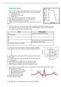
1. Figure 1 shows a typical action potential that occurs in certain cardiac
cells. Which of the following answers matches this action potential?
a. Autorhythmic cells
b. Myocardial contractile cells
c. Neuron cells
1b. Draw each of the earlier mentioned cells action potential.
1c. What phases are labeled with A, B, C, D and E in figure 1?
1d. What is the function of phase C in figure 1?
1e. What period of the action potential will be longer due to phase C in
figure 1?
2. When the heart beats, certain mechanics make it possible to pump blood through the vessels.
Each heartbeat can be seen as one cardiac cycle. The following table consists out of 5 phases that
occur during one cardiac cycle. Complete the table.
Phase What happens?
1 Chambers relaxes and fill passively
2 Atrial systole
3
4 Pressure rises due to contraction and causing
the semilunar valves to close
5 Isovolumic ventricular relaxation
2b. The cardiac cycle can be pictured as a loop in a graphic, see figure 2.
Use the phases in the previous table to complete the following answers.
A=…
B=…
C=…
D=…
2c. On what point occurs the end-systolic volume (ESV), mark the point
on the graphic.
2d. On what point occurs the end-diastolic volume (EDV), mark the
point on the graphic.
2e. What happens to the graphic when the preload increases?
2f. What happens to the graphic when the afterload increases?
2g. What happens to the graphic when the contractility increases?
3. ECG stands for electrocardiogram and can be divided into P-wave, QRS-complex and T-wave.
What causes the P-waves?
a. Depolarization of the ventricles
b. Repolarization of the ventricles
c. Depolarization of the atria
d. Repolarization of the atria
3b. What causes the QRS-complex?
3c. What causes the T-wave?
25 Q’s of BBS1002 – for a full summary, try out summary of BBS1002