THEME 1: NEOPLASIA
DERMATOPATHOLOGY
NEOPLASIA
− neoplasm = genetically driven growth of cells → benign, pre-malignant, malignant
− tumour:
o always a formation of mass
o can be genetically driven → neoplasm
o can be due to other causes e.g., inflammation
− neoplasm → isn’t always a mass e.g., leukaemia
− benign neoplasm:
o slow and local growth
o no metastases
o no atypia/anaplasia (stop looking like precursors)
o moles, wards (bradavice)
o isn’t always premalignant
− malignant neoplasm:
o often rapid growth/proliferation (mitosis in tissues)
o infiltration in other tissues → metastasizing
o atypical cells → dysplasia = abnormal
development of cells within tissues or organs
− multiple genetic aberrations in different genes → usually
at least 6 genes need to be hit:
1. autonomous cell proliferation → growth
2. new vessels formation → growth
3. loss of cell apoptosis → growth, survival
4. loss of differentiation → loss of function
5. loss of cell contact inhibition → invasion
6. growth in and out of the vessels → metastasis
→ different mutations need to occur for each step
→ every new hit leads towards malignancy
→ also keeps happening within tumours, however, it doesn’t spread
− cancer genes:
o (proto)oncogenes = code for proteins promoting for cell growth, gain of function mutation
(activation or gain of expression)
o tumour suppressor genes = code for proteins inhibiting cell proliferation or inducing cell
apoptosis, loss of function mutation (loss of gene or loss of expression) → needs to mutate
in both alleles
o DNA repair genes = code for proteins controlling and repairing the genome, loss of function
mutation (also 2x) → accumulation of DNA errors
o viral genes = incorporated into human genes and cause cancer
,− genetic alterations:
o mutations:
▪ small
▪ substitution = activation of oncogenes → hotspot mutation = a continuous
activation of the gene & inhibition of tumour suppressor → stop codon
▪ insertion/deletion = + frame shift → inhibition of tumour suppressor & - frameshift
→ gain or loss of function
o copy number variations:
▪ large
▪ sections of the genome are repetead in a
chromosome
▪ gain of oncogenes
▪ loss of tumour suppressor genes
o chromosomal translocations:
▪ expression/activation of oncogenes
▪ part of a chromosome is fused to another
chromosome → fusion of 2 genes
− somatic mutation:
o acquired
o only in certain cells of the body
o dependent on a carcinogen
− germline mutations:
o often in the family history
o congenital (urođeno)
o present in all cells of the body
o can give the susceptibility for cancer
CLINICAL PATHOLOGY
− clinical pathologist = a medical specialist who diagnoses disease by investigating tissues (and cells)
of patients
− pathological investigation of skin tumours:
1. assessment of the differentiation line → from which tissue/cells the tumour derived
2. establishing whether the tumour is benign or malignant
1 + 2 = diagnosis
3. determine the tumour stage → measuring invasion depth or looking at which tissues it invaded
→ dyeing the margins before cutting
4. establishing whether the margins are free of tumour
→ determines clinical management: tumour size, necessity of (re)excision and its margins, necessity of
adjuvant radiotherapy, node excision, and adjuvant systemic therapy
,− example:
− pathological examination of skin excision:
o skin excision with marked margins
o cut with bread loaf technique
o representative material goes under
the microscope → the thinner slices
− diagnosis:
o routine histology = sufficient in most
cases, H&E (haematoxylin and eosin)
staining, H/purple – nucleus & E/pink
– cytoplasm
o immunohistochemistry = usage of an antibody directed against an antigen only expressed
on tumour cells, the binding is visualized by the 2nd antibody with an enzyme which then
converts a chromogen into an insoluble stain on the section, used for determining the
tumour/cell origin and for the malignant character of the tumour
o molecular pathology → used to make diagnosis and for determining treatment options
SKIN TUMOURS
− the largest organ, aging, lifelong exposure to UV light, tanning bed → increase of skin cancer
− every cancer cause has its own mutational signature
− other causes: viral infection (HPV), hereditary
− different skin structures: epidermis, dermis, hair follicles, oil and sweat glands, nerves, fatty tissue,
lymph vessels, melanin cells, immune cells → all can give rise to different tumours
− basal cell carcinoma/BCC:
o most frequent skin cancer → becoming an epidemic
o derived from hair follicles
o clinical behaviour: locally aggressive, can be mutilating, however, hardly ever metastizes
o location: always present in the skin, frequent around the eyes and the nose, not found on
mucosa, palms, and soles → similar to hair follicles
, o histology: large dark nuclei with very
little cytoplasm
o etiology (the cause of a disease or an
abnormal condition): UV-specific
mutation, age > 40, only Caucasians,
areas with cumulative sun-
exposition (face and hands)
− squamous cell carcinoma/SCC:
o common and rising
o derived from keratinocytes (major
cell type of the epidermis)
o mostly in areas with cumulative
sun-exposition
o mutilating, but only 2% of them
can metastize
o histology: always show
keratinization (forming of keratin,
dying skin cells)
o etiology: UVB → tumours develop
on the sun-exposed sites, UV-
specific mutation & HPV → larger
cells with better margins and
irregular nuclei, hypertrophic
epidermis, no invasive growth,
especially in immune compromised patients
o 2 types of dysplasia: actinic keratosis (UVB) & Morbus Bowen (HPV)
MELANOCYTIC TUMOURS
− melanocyte:
o pigment producing cell
o in the basal layer of the epidermis
between keratinocytes
o 1 melanocyte communicates with
30-40 keratinocytes via dendrites
o melanin granules protect
keratinocytes against UV
o darker skin → more melanin
produced, same number of
melanocytes
BENIGN MELANOCYTIC TUMOURS → NEVUS (MADEŽ)
− develop mostly in the first 2 years of life
− most people have 20-30 nevi
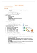
− neoplasms → genetically driven → 60% BRAF gene & 20% NRAS gene mutations

